Anti-Caspase-3 antibody (ab4051)
Key features and details
- Rabbit polyclonal to Caspase-3
- Suitable for: WB, IHC-P
- Reacts with: Human
- Isotype: IgG
Overview
-
Product name
Anti-Caspase-3 antibody
See all Caspase-3 primary antibodies -
Description
Rabbit polyclonal to Caspase-3 -
Host species
Rabbit -
Specificity
Caspase-3 is a member of the interleukin-1 β-converting enzyme family. Caspase-3 is thought to be associated with induction of apoptosis. Caspase-3 is synthesized as inactive 32 kDa proenzyme and is processed during apoptosis generating two subunits of 17 kDa and 12 kDa. Caspase-3 stains the epithelial cells of skin, renal proximal tubules and collecting ducts.
This antibody reacts with the 32 kDa proenzyme and the 17 kDa active form of caspase 3.
-
Tested applications
Suitable for: WB, IHC-Pmore details -
Species reactivity
Reacts with: Human -
Immunogen
Synthetic peptide corresponding to Human Caspase-3 aa 167-175. Corresponding to the cleavage site of human caspase 3.
Database link: P42574
Properties
-
Form
Liquid -
Storage instructions
Shipped at 4°C. Store at +4°C short term (1-2 weeks). Upon delivery aliquot. Store at -20°C. Avoid freeze / thaw cycle. -
Storage buffer
Preservative: 0.05% Sodium azide
Constituent: 1% BSA -
 Concentration information loading...
 -
Purity
IgG fraction -
Purification notes
Purified immunoglobulin fraction of rabbit antiserum against Caspase-3 containing sodium azide as a preservative. -
Clonality
Polyclonal -
Isotype
IgG -
Research areas
Images
-
 Immunohistochemistry (Formalin/PFA-fixed paraffin-embedded sections) - Anti-Caspase-3 antibody (ab4051)
ab4051 staining Caspase-3 in formalin-fixed, paraffin-embedded Human tonsil tissue by Immunohistochemistry.
-
Anti-Caspase-3 antibody (ab4051) + Human Lung Cell Extract
Predicted band size: 31 kDa
-
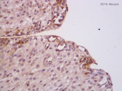
 Immunohistochemistry (Formalin/PFA-fixed paraffin-embedded sections) - Anti-Caspase-3 antibody (ab4051) Image courtesy of an anonymous AbReview
ab4051 staining Caspase 3 in Pig liver tissue sections by Immunohistochemistry (IHC-P - paraformaldehyde-fixed, paraffin-embedded sections). Tissue was fixed with formaldehyde and blocked with 5% serum for 1 hour at 21°C; antigen retrieval was by heat mediation in a citrate buffer. Samples were incubated with primary antibody (1/100 in milk) for 20 hours at 4°C. A Biotin-conjugated Goat anti-rabbit IgG polyclonal (1/500) was used as the secondary antibody.
-
 Immunohistochemistry (Formalin/PFA-fixed paraffin-embedded sections) - Anti-Caspase-3 antibody (ab4051) Image courtesy of an anonymous AbReview
ab4051 staining Caspase 3 in Cow Ovary tissue sections by Immunohistochemistry (IHC-P - paraformaldehyde-fixed, paraffin-embedded sections). Tissue was fixed with formaldehyde and blocked with 10% serum for 15 minutes at 25°C; antigen retrieval was by heat mediation in a citrate buffer. Samples were incubated with primary antibody (1/75 in PBS + BSA) for 16 hours at 4°C. A Biotin-conjugated Goat anti-rabbit IgG polyclonal (1/100) was used as the secondary antibody.


