Anti-HMGB2 antibody (ab113929)
")
Key features and details
- Rabbit polyclonal to HMGB2
- Suitable for: IHC-P, ICC/IF, WB
- Knockout validated
- Reacts with: Human
- Isotype: IgG
Overview
-
Product name
Anti-HMGB2 antibody
See all HMGB2 primary antibodies -
Description
Rabbit polyclonal to HMGB2 -
Host species
Rabbit -
Tested Applications & Species
See all applications and species dataApplication Species ICC/IF HumanIHC-P HumanWB Human
-
Immunogen
Synthetic peptide. This information is proprietary to Abcam and/or its suppliers.
-
Positive control
- This antibody gave a positive signal in HeLa Nuclear lysate as well as the following whole cell lysates: HepG2; U20S; A549; MCF7; K562. This antibody gave a positive result in IF in the following methanol fixed cell lines: HepG2 It also gave a positive result in FFPE human kidney RCC tissue sections
Properties
-
Form
Liquid -
Storage instructions
Shipped at 4°C. Store at +4°C short term (1-2 weeks). Upon delivery aliquot. Store at -20°C or -80°C. Avoid freeze / thaw cycle. -
Storage buffer
pH: 7.40
Preservative: 0.02% Sodium azide
Constituent: PBS
Batches of this product that have a concentration Concentration information loading...
Purity
Immunogen affinity purifiedClonality
PolyclonalIsotype
IgGResearch areas
Associated products
-
Compatible Secondaries
-
Isotype control
-
Recombinant Protein
Applications
The Abpromise guarantee
Our Abpromise guarantee covers the use of ab113929 in the following tested applications.
The application notes include recommended starting dilutions; optimal dilutions/concentrations should be determined by the end user.
GuaranteedTested applications are guaranteed to work and covered by our Abpromise guarantee.
PredictedPredicted to work for this combination of applications and species but not guaranteed.
IncompatibleDoes not work for this combination of applications and species.
Application Species ICC/IF HumanIHC-P HumanWB HumanAll applications MouseRabbitHorseCowDogChimpanzeeMacaque monkeyGorillaChinese hamsterOrangutanApplication Abreviews Notes IHC-P Use a concentration of 5 µg/ml. Perform heat mediated antigen retrieval before commencing with IHC staining protocol.ICC/IF Use a concentration of 5 µg/ml.WB Use a concentration of 1 µg/ml. Detects a band of approximately 28 kDa (predicted molecular weight: 24 kDa).Notes IHC-P
Use a concentration of 5 µg/ml. Perform heat mediated antigen retrieval before commencing with IHC staining protocol.ICC/IF
Use a concentration of 5 µg/ml.WB
Use a concentration of 1 µg/ml. Detects a band of approximately 28 kDa (predicted molecular weight: 24 kDa).Target
-
Function
DNA binding proteins that associates with chromatin and has the ability to bend DNA. Binds preferentially single-stranded DNA. Involved in V(D)J recombination by acting as a cofactor of the RAG complex. Acts by stimulating cleavage and RAG protein binding at the 23 bp spacer of conserved recombination signal sequences (RSS). -
Sequence similarities
Belongs to the HMGB family.
Contains 2 HMG box DNA-binding domains. -
Cellular localization
Nucleus. Chromosome. - Information by UniProt
-
Database links
- Entrez Gene: 540444 Cow
- Entrez Gene: 3148 Human
- Entrez Gene: 97165 Mouse
- Omim: 163906 Human
- SwissProt: P40673 Cow
- SwissProt: P26583 Human
- SwissProt: P30681 Mouse
- Unigene: 434953 Human
see all -
Alternative names
- C80539 antibody
- High mobility group (nonhistone chromosomal) protein 2 antibody
- High mobility group box 2 antibody
see all
Images
-
Western blot - Anti-HMGB2 antibody (ab113929)All lanes : Anti-HMGB2 antibody (ab113929) at 1 µg/ml
Lane 1 : Wild-type HAP1 whole cell lysate
Lane 2 : HMGB2 knockout HAP1 whole cell lysate
Lane 3 : HeLa whole cell lysate
Lane 4 : K562 whole cell lysate
Lysates/proteins at 20 µg per lane.
Predicted band size: 24 kDaLanes 1 - 4: Merged signal (red and green). Green - ab113929 observed at 30 kDa. Red - loading control, ab9484, observed at 37 kDa.
ab113929 was shown to specifically react with HMGB2 in wild-type HAP1 cells as signal was lost in HMGB2 knockout cells. Wild-type and HMGB2 knockout samples were subjected to SDS-PAGE. Ab113929 and ab9484 (Mouse anti-GAPDH loading control) were incubated overnight at 4°C at 1 μg/ml and 1/20000 dilution respectively. Blots were developed with Goat anti-Rabbit IgG H&L (IRDye® 800CW) preabsorbed ab216773 and Goat anti-Mouse IgG H&L (IRDye® 680RD) preabsorbed ab216776 secondary antibodies at 1/20000 dilution for 1 hour at room temperature before imaging. -
 Western blot - Anti-HMGB2 antibody (ab113929)All lanes : Anti-HMGB2 antibody (ab113929) at 1 µg/ml
Lane 1 : HepG2 (Human hepatocellular liver carcinoma cell line) Whole Cell Lysate
Lane 2 : U2OS (Human osteosarcoma cell line) Whole Cell Lysate
Lane 3 : A549 (Human lung adenocarcinoma epithelial cell line) Whole Cell Lysate
Lane 4 : MCF7 (Human breast adenocarcinoma cell line) Whole Cell Lysate
Lane 5 : HeLa (Human epithelial carcinoma cell line) Nuclear Lysate
Lane 6 : K562 (Human erythromyeloblastoid leukemia cell line) Whole Cell Lysate
Lysates/proteins at 10 µg per lane.
Secondary
All lanes : Goat Anti-Rabbit IgG H&L (HRP) preadsorbed (ab97080) at 1/5000 dilution
Developed using the ECL technique.
Performed under reducing conditions.
Predicted band size: 24 kDa
Observed band size: 28 kDa why is the actual band size different from the predicted?
Additional bands at: 100 kDa, 300 kDa, 50 kDa. We are unsure as to the identity of these extra bands.
Exposure time: 8 minutes -
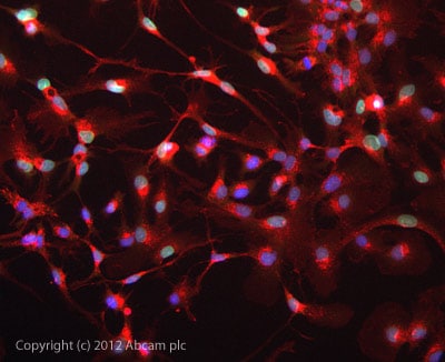 Immunocytochemistry/ Immunofluorescence - Anti-HMGB2 antibody (ab113929)
ICC/IF image of ab113929 stained HepG2 cells. The cells were 100% methanol fixed (5 min) and then incubated in 1%BSA / 10% normal goat serum / 0.3M glycine in 0.1% PBS-Tween for 1h to permeabilise the cells and block non-specific protein-protein interactions. The cells were then incubated with the antibody ab113929 at 5µg/ml overnight at +4°C. The secondary antibody (green) was DyLight® 488 goat anti- rabbit (ab96899) IgG (H+L) used at a 1/1000 dilution for 1h. Alexa Fluor® 594 WGA was used to label plasma membranes (red) at a 1/200 dilution for 1h. DAPI was used to stain the cell nuclei (blue) at a concentration of 1.43µM.
-
 Immunohistochemistry (Formalin/PFA-fixed paraffin-embedded sections) - Anti-HMGB2 antibody (ab113929)IHC image of ab113929 staining in human kidney RCC formalin fixed paraffin embedded tissue section, performed on a Leica BondTM system using the standard protocol F. The section was pre-treated using heat mediated antigen retrieval with sodium citrate buffer (pH6, epitope retrieval solution 1) for 20 mins. The section was then incubated with ab113929, 5µg/ml, for 15 mins at room temperature and detected using an HRP conjugated compact polymer system. DAB was used as the chromogen. The section was then counterstained with haematoxylin and mounted with DPX.
For other IHC staining systems (automated and non-automated) customers should optimize variable parameters such as antigen retrieval conditions, primary antibody concentration and antibody incubation times.
Protocols
Datasheets and documents
References (0)
ab113929 has not yet been referenced specifically in any publications.
Images
-
Western blot - Anti-HMGB2 antibody (ab113929)All lanes : Anti-HMGB2 antibody (ab113929) at 1 µg/ml
Lane 1 : Wild-type HAP1 whole cell lysate
Lane 2 : HMGB2 knockout HAP1 whole cell lysate
Lane 3 : HeLa whole cell lysate
Lane 4 : K562 whole cell lysate
Lysates/proteins at 20 µg per lane.
Predicted band size: 24 kDaLanes 1 - 4: Merged signal (red and green). Green - ab113929 observed at 30 kDa. Red - loading control, ab9484, observed at 37 kDa.
ab113929 was shown to specifically react with HMGB2 in wild-type HAP1 cells as signal was lost in HMGB2 knockout cells. Wild-type and HMGB2 knockout samples were subjected to SDS-PAGE. Ab113929 and ab9484 (Mouse anti-GAPDH loading control) were incubated overnight at 4°C at 1 μg/ml and 1/20000 dilution respectively. Blots were developed with Goat anti-Rabbit IgG H&L (IRDye® 800CW) preabsorbed ab216773 and Goat anti-Mouse IgG H&L (IRDye® 680RD) preabsorbed ab216776 secondary antibodies at 1/20000 dilution for 1 hour at room temperature before imaging. -
Western blot - Anti-HMGB2 antibody (ab113929)All lanes : Anti-HMGB2 antibody (ab113929) at 1 µg/ml
Lane 1 : HepG2 (Human hepatocellular liver carcinoma cell line) Whole Cell Lysate
Lane 2 : U2OS (Human osteosarcoma cell line) Whole Cell Lysate
Lane 3 : A549 (Human lung adenocarcinoma epithelial cell line) Whole Cell Lysate
Lane 4 : MCF7 (Human breast adenocarcinoma cell line) Whole Cell Lysate
Lane 5 : HeLa (Human epithelial carcinoma cell line) Nuclear Lysate
Lane 6 : K562 (Human erythromyeloblastoid leukemia cell line) Whole Cell Lysate
Lysates/proteins at 10 µg per lane.
Secondary
All lanes : Goat Anti-Rabbit IgG H&L (HRP) preadsorbed (ab97080) at 1/5000 dilution
Developed using the ECL technique.
Performed under reducing conditions.
Predicted band size: 24 kDa
Observed band size: 28 kDa why is the actual band size different from the predicted?
Additional bands at: 100 kDa, 300 kDa, 50 kDa. We are unsure as to the identity of these extra bands.
Exposure time: 8 minutes
-
Immunocytochemistry/ Immunofluorescence - Anti-HMGB2 antibody (ab113929)
ICC/IF image of ab113929 stained HepG2 cells. The cells were 100% methanol fixed (5 min) and then incubated in 1%BSA / 10% normal goat serum / 0.3M glycine in 0.1% PBS-Tween for 1h to permeabilise the cells and block non-specific protein-protein interactions. The cells were then incubated with the antibody ab113929 at 5µg/ml overnight at +4°C. The secondary antibody (green) was DyLight® 488 goat anti- rabbit (ab96899) IgG (H+L) used at a 1/1000 dilution for 1h. Alexa Fluor® 594 WGA was used to label plasma membranes (red) at a 1/200 dilution for 1h. DAPI was used to stain the cell nuclei (blue) at a concentration of 1.43µM.
-
Immunohistochemistry (Formalin/PFA-fixed paraffin-embedded sections) - Anti-HMGB2 antibody (ab113929)IHC image of ab113929 staining in human kidney RCC formalin fixed paraffin embedded tissue section, performed on a Leica BondTM system using the standard protocol F. The section was pre-treated using heat mediated antigen retrieval with sodium citrate buffer (pH6, epitope retrieval solution 1) for 20 mins. The section was then incubated with ab113929, 5µg/ml, for 15 mins at room temperature and detected using an HRP conjugated compact polymer system. DAB was used as the chromogen. The section was then counterstained with haematoxylin and mounted with DPX.
For other IHC staining systems (automated and non-automated) customers should optimize variable parameters such as antigen retrieval conditions, primary antibody concentration and antibody incubation times.


