Anti-SOX2 antibody (ab97959)
Key features and details
- Rabbit polyclonal to SOX2
- Suitable for: ICC, IHC-P, WB
- Reacts with: Mouse, Rat, Human
- Isotype: IgG
Overview
-
Product name
Anti-SOX2 antibody
See all SOX2 primary antibodies -
Description
Rabbit polyclonal to SOX2 -
Host species
Rabbit -
Tested Applications & Species
See all applications and species dataApplication Species ICC MouseRatHumanIHC-FrFl HumanWB MouseRatHuman -
Immunogen
Synthetic peptide. This information is proprietary to Abcam and/or its suppliers.
-
Positive control
- ICC: NCCIT and NIH/3T3 cells. Dissociated induced pluripotent stem cells from mouse embryonic fibroblasts. Mouse embryonic stem cells. WB: NCCIT, IOUD2, HUES7, F9 and MCF7 whole cell lysate. IHC:Human brain glioma.
-
General notes
Images
-
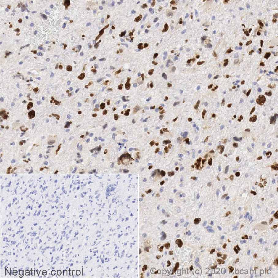
IHC image of SOX2 staining in Human brain glioma formalin fixed paraffin embedded tissue section, performed on a Leica Bond™ system using the standard protocol F. The section was pre-treated using heat mediated antigen retrieval with sodium citrate buffer (pH6, epitope retrieval solution 1) for 20 mins. The section was then incubated with ab97959, 1µg/ml, for 15 mins at room temperature and detected using an HRP conjugated compact polymer system. DAB was used as the chromogen. The section was then counterstained with haematoxylin and mounted with DPX.
For other IHC staining systems (automated and non-automated) customers should optimize variable parameters such as antigen retrieval conditions, primary antibody concentration and antibody incubation times.
-
ab97959 staining SOX2 in primary hippocampal rat neurons/glia, (obtained from Neuromics, cat. no. PC35101), DIV14. The cells were fixed with 4% paraformaldehyde (10 min), permeabilized with 0.1% PBS-Triton X-100 for 5 minutes and then blocked with 1% BSA/10% normal goat serum/0.3M glycine in 0.1%PBS-Tween for 1h. The cells were then incubated overnight at 4°C with ab97959 at 0.1µg/ml and ab7291, Mouse monoclonal [DM1A] to alpha Tubulin - Loading Control. Cells were then incubated with ab150081, Goat polyclonal Secondary Antibody to Rabbit IgG - H&L (Alexa Fluor® 488), pre-adsorbed at 1/1000 dilution (shown in green) and ab150120, Goat polyclonal Secondary Antibody to Mouse IgG - H&L (Alexa Fluor® 594), pre-adsorbed at 1/1000 dilution (shown in pseudocolour red). Nuclear DNA was labelled with DAPI (shown in blue).
Image was acquired with a high-content analyser (Operetta CLS, Perkin Elmer) and a maximum intensity projection of confocal sections is shown.
-
Cell line: NCCIT (human pluripotent embryonal carcinoma)
Target AbID: Ab97959 anti-Sox2, Goat Anti-Rabbit IgG H&L (Alexa Fluor® 488) (ab150077) secondary antibody was used.
Counterstain AbID: Ab7291 anti-Tubulin (Rabbit mAb), 97959
Fixative: 4% PFA
Permeabilisation: 0.1% Triton-X
Nuclear counter stain: DAPI
Comments: Confocal image showing negative staining on NCCIT cells
Target primary antibody dilution: 1:500
Target secondary antibody dilution: 1:1000 (2ug/mL)
Counterstain primary antibody dilution: 1:1000 (1ug/mL)
Counterstain secondary antibody dilution: 1:1000 (2ug/mL)
Negative control 1 primary antibody dilution: 1:500 (Ab97959)
Negative control 1 secondary antibody dilution: 1:1000 (2ug/mL) (Ab150120)
Negative control 2 primary antibody dilution: 1:1000 (1ug/mL) (Ab7291)
Negative control 2 secondary antibody dilution: 1:1000 (2ug/mL) (Ab150077)
-
All lanes : Anti-SOX2 antibody (ab97959) at 1 µg
Lane 1 : NCCIT (Human embryonic carcinoma cell line) Whole Cell Lysate
Lane 2 : F9 (Mouse embryonic carcinoma cell line) Whole Cell Lysate
Lane 3 : MCF7 (Human breast adenocarcinoma cell line) Whole Cell Lysate
Lane 4 : C6 (Rat glial tumor cell line) Whole Cell Lysate
Lane 5 : Hippocampus (Rat) Tissue Lysate
Lane 6 : Spinal Cord (Rat) Tissue Lysate
Lysates/proteins at 10 µg per lane.
Secondary
All lanes : Blots were developed with goat anti-rabbit IgG (H+L) and goat anti-mouse IgG (H+L) secondary antibodies at 1/10,000 dilution for 1 h at room temperature before imaging.
Performed under reducing conditions.
Predicted band size: 34 kDa
Observed band size: 40 kDa why is the actual band size different from the predicted?Green signal from target - ab97959 observed at 40 kDa
Red signal from loading control ab9484 (GAPDH) observed at 37 kDa -
Cell line: NIH/3T3 (mouse embryonic fibroblast cell line)
Target AbID: Ab97959 anti-Sox2, used Goat Anti-Rabbit IgG H&L (Alexa Fluor® 488) (ab150077) secondary antibody
Counterstain AbID: Ab7291 anti-Tubulin (Rabbit mAb), Ab150120 AlexaFluor®594 Goat anti-Mouse secondary
Fixative: 4% PFA
Permeabilisation: 0.1% Triton-X
Nuclear counter stain: DAPI
Comments: Confocal image showing negative staining on NIH/3T3 cells
Target primary antibody dilution: 1:500
Target secondary antibody dilution: 1:1000 (2ug/mL)
Counterstain primary antibody dilution: 1:1000 (1ug/mL)
Counterstain secondary antibody dilution: 1:1000 (2ug/mL)
Negative control 1 primary antibody dilution: 1:500 (Ab97959)
Negative control 1 secondary antibody dilution: 1:1000 (2ug/mL) (Ab150120)
Negative control 2 primary antibody dilution: 1:1000 (1ug/mL) (Ab7291)
Negative control 2 secondary antibody dilution: 1:1000 (2ug/mL) (Ab150077)
-
 Immunocytochemistry - Anti-SOX2 antibody (ab97959) Muratore CR et al. Comparison and Optimization of hiPSC Forebrain Cortical Differentiation Protocols. PLoS One 9:e105807 (2014). Reproduced under the Creative Commons license http://creativecommons.org/licenses/by/4.0/
ICC/IF analysis of dissociated induced pluripotent stem cells from mouse embryonic fibroblasts stained for SOX-2 (Red) using ab97959. TOPRO (purple). Scale bar=100 μm
-
ICC/IF image of ab97959 stained mouse embryonic stem cells. The cells were 4% formaldehyde fixed (10 min) and then incubated in 1%BSA / 10% normal goat serum / 0.3M glycine in 0.1% PBS-Tween for 1h to permeabilise the cells and block non-specific protein-protein interactions. The cells were then incubated with the antibody (ab97959, 1µg/ml) overnight at +4°C. The secondary antibody (green) was Alexa Fluor® 488 goat anti-rabbit IgG (H+L) used at a 1/1000 dilution for 1h. Alexa Fluor® 594 WGA was used to label plasma membranes (red) at a 1/200 dilution for 1h. DAPI was used to stain the cell nuclei (blue) at a concentration of 1.43µM.
-
All lanes : Anti-SOX2 antibody (ab97959) at 1 µg/ml
Lane 1 : IOUD2 (Mouse embryonic stem cell) Whole Cell Lysate
Lane 2 : HUES7 (Human embryonic stem cell line) Whole Cell Lysate
Lysates/proteins at 10 µg per lane.
Secondary
All lanes : Goat polyclonal to Rabbit IgG - H&L - Pre-Adsorbed (HRP) at 1/3000 dilution
Developed using the ECL technique.
Performed under reducing conditions.
Predicted band size: 34 kDa
Observed band size: 43 kDa why is the actual band size different from the predicted?
Additional bands at: 37 kDa, 39 kDa. We are unsure as to the identity of these extra bands.
Exposure time: 3 minutes


