Anti-Cofilin (phospho S3) antibody (ab12866)
 antibody (ab12866)")
Key features and details
- Rabbit polyclonal to Cofilin (phospho S3)
- Suitable for: ICC/IF, WB, Flow Cyt
- Reacts with: Mouse, Rat, Human, African green monkey
- Isotype: IgG
Overview
-
Product name
Anti-Cofilin (phospho S3) antibody
See all Cofilin primary antibodies -
Description
Rabbit polyclonal to Cofilin (phospho S3) -
Host species
Rabbit -
Tested Applications & Species
See all applications and species dataApplication Species Flow Cyt HumanICC/IF HumanWB Human
-
Immunogen
Synthetic peptide derived from a region of human cofilin that contains serine 3. The sequence is conserved in mouse and rat.
-
General notes
The Life Science industry has been in the grips of a reproducibility crisis for a number of years. Abcam is leading the way in addressing the problem with our range of recombinant monoclonal antibodies and knockout edited cell lines for gold-standard validation.
One factor contributing to the crisis is the use of antibodies that are not suitable. This can lead to misleading results and the use of incorrect data informing project assumptions and direction. To help address this challenge, we have introduced an application and species grid on our primary antibody datasheets to make it easy to simplify identification of the right antibody for your needs.
Learn more here.
Properties
-
Form
Liquid -
Storage instructions
Shipped at 4°C. Upon delivery aliquot and store at -20°C. Avoid freeze / thaw cycles. -
Storage buffer
pH: 7.3
Preservative: 0.05% Sodium azide
Constituents: PBS, 50% Glycerol, 0.1% BSA
PBS (without Mg2+ and Ca2+), BSA (IgG, protease free) -
 Concentration information loading...
 -
Purity
Immunogen affinity purified -
Clonality
Polyclonal -
Isotype
IgG -
Research areas
Images
-
Western blot - Anti-Cofilin (phospho S3) antibody (ab12866)Peptide Competition and Phosphatase Treatment
Lysates prepared from MDCK cells treated with staurosporine (1) or left untreated (2-6) were resolved by SDS-PAGE on a 10% polyacrylamide gel and transferred to PVDF. Membranes were either left untreated (1-5) or treated with lambda phosphatase (6), blocked with a 5% BSA-TBST buffer for one hour at room temperature, and incubated with the ab12866 antibody for two hours at room temperature in a 3% BSA-TBST buffer, following prior incubation with: no peptide (1, 2, 6), the non phosphopeptide corresponding to the immunogen (3), a generic phosphoserine-containing peptide (4) or, the phosphopeptide immunogen (5). After washing, membranes were incubated with goat F(ab)2 anti-rabbit IgG HRP conjugate and bands were detected using the Pierce SuperSignal method. The data show that only the peptide corresponding to cofilin [pS3] blocks the antibody signal. The data also show that phosphatase stripping eliminates the signal, verifying that the anti -
 Immunocytochemistry/ Immunofluorescence - Anti-Cofilin (phospho S3) antibody (ab12866)ICC/IF image of ab12866 stained MCF7 cells. The cells were 4% PFA fixed (10 min) and then incubated in 1%BSA / 10% normal goat serum / 0.3M glycine in 0.1% PBS-Tween for 1h to permeabilise the cells and block non-specific protein-protein interactions. The cells were then incubated with the antibody (ab12866, 1µg/ml) overnight at +4°C. The secondary antibody (green) was Alexa Fluor® 488 goat anti-rabbit IgG (H+L) used at a 1/1000 dilution for 1h. Alexa Fluor® 594 WGA was used to label plasma membranes (red) at a 1/200 dilution for 1h. DAPI was used to stain the cell nuclei (blue) at a concentration of 1.43µM.
-
 Immunocytochemistry/ Immunofluorescence - Anti-Cofilin (phospho S3) antibody (ab12866) This image is courtesy of an anonymous abreview.
Immunocytochemistry/ Immunofluorescence analysis of human neuroblastoma cells labeling Cofilin (phospho S3) with ab12866 at 1/500 dilution. Cells were fixed in paraformaldehyde, permeabilized for 15 minutes in 0.01% Triton X-100, blocked using 5% serum for 30 minutes at 20°C, then incubated with ab12866 at a 1/500 dilution for 2 hours at 20°C. The secondary used was a Dylight 488 conjugated donkey anti-rabbit IgG (H+L) used at a 1/500 dilution.
-
 Western blot - Anti-Cofilin (phospho S3) antibody (ab12866)All lanes : Anti-Cofilin (phospho S3) antibody (ab12866) at 1 µg/ml
Lane 1 : HeLa whole cell extract
Lane 2 : HeLa treated for overnight with 150 uM of H2O2 whole cell extract
Lane 3 : HeLa treated for overnight with 3 uM of Staurosporine whole cell extract
Lane 4 : COS-7 whole cell extract
Lane 5 : NIH/3T3 (Mouse embryo fibroblast cell line) whole cell extract
Lane 6 : MCF7 whole cell extract
Lane 7 : Rat Skeletal Muscle whole cell extract
Lane 8 : A-431 whole cell extract
Lane 9 : A-431 treated for overnight with 150 uM of H2O2 whole cell extract
Lane 10 : Jurkat whole cell extract
Lane 11 : Jurkat treated for overnight with 3 uM of Staurosporine whole cell extract
Lysates/proteins at 20 µg per lane.
Secondary
All lanes : Goat anti-rabbit IgG (H+L), HRP conjugate at 1/2500 dilution
Observed band size: 18 kDa why is the actual band size different from the predicted?
-
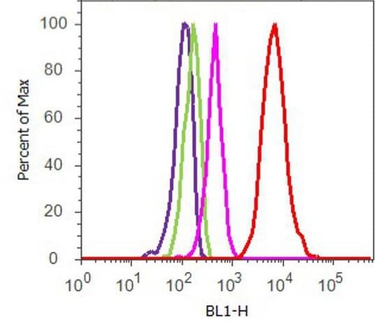 Flow Cytometry - Anti-Cofilin (phospho S3) antibody (ab12866)
Flow Cytometry analysis of U-87 MG cells labeling Cofilin (phospho S3) with ab12866. Cells were fixed with 70% ethanol for 10 minutes, permeabilized with 0.25% Triton™ X-100 for 20 minutes, and blocked with 5% BSA for 30 minutes at room temperature. Cells were labeled with Anti-Cofilin (phospho S3) antibody (ab12866, red) or with rabbit isotype control (pink) at 3-5 ug/million cells in 2.5% BSA. After incubation at room temperature for 2 hours, the cells were labeled with Alexa Fluor® 488 Goat Anti-Rabbit Secondary Antibody at a dilution of 1/400 for 30 minutes at room temperature. The representative 10,000 cells were acquired and analyzed for each sample. The purple histogram represents unstained control cells and the green histogram represents no-primary-antibody control.


