Anti-p95/NBS1 antibody (ab23996)
Key features and details
- Rabbit polyclonal to p95/NBS1
- Suitable for: WB, ICC/IF
- Reacts with: Mouse, Human
- Isotype: IgG
Overview
-
Product name
Anti-p95/NBS1 antibody
See all p95/NBS1 primary antibodies -
Description
Rabbit polyclonal to p95/NBS1 -
Host species
Rabbit -
Tested applications
Suitable for: WB, ICC/IFmore details -
Species reactivity
Reacts with: Mouse, Human
Predicted to work with: Rat, Chicken, Dog
-
Immunogen
Synthetic peptide corresponding to Human p95/NBS1 aa 700 to the C-terminus (C terminal) conjugated to keyhole limpet haemocyanin.
(Peptide available asab24289) -
Positive control
- ab23996 gave a positive result in the following lysates: MRC5 whole cell lysate (SV40 transformed immortal (WT) fibroblasts), Hela whole cell, NIH3T3 whole cell, HepG2 whole cell and Jurkat whole cell.
-
General notes
This product was previously labelled as p95 NBS1
Reproducibility is key to advancing scientific discovery and accelerating scientists’ next breakthrough.
Abcam is leading the way with our range of recombinant antibodies, knockout-validated antibodies and knockout cell lines, all of which support improved reproducibility.
We are also planning to innovate the way in which we present recommended applications and species on our product datasheets, so that only applications & species that have been tested in our own labs, our suppliers or by selected trusted collaborators are covered by our Abpromise™ guarantee.
In preparation for this, we have started to update the applications & species that this product is Abpromise guaranteed for.
We are also updating the applications & species that this product has been “predicted to work with,” however this information is not covered by our Abpromise guarantee.
Applications & species from publications and Abreviews that have not been tested in our own labs or in those of our suppliers are not covered by the Abpromise guarantee.
Please check that this product meets your needs before purchasing. If you have any questions, special requirements or concerns, please send us an inquiry and/or contact our Support team ahead of purchase. Recommended alternatives for this product can be found below, as well as customer reviews and Q&As.
Properties
-
Form
Liquid -
Storage instructions
Shipped at 4°C. Store at +4°C short term (1-2 weeks). Upon delivery aliquot. Store at -20°C or -80°C. Avoid freeze / thaw cycle. -
Storage buffer
pH: 7.40
Preservative: 0.02% Sodium azide
Constituent: PBS
Batches of this product that have a concentration Concentration information loading...
Purity
Immunogen affinity purifiedClonality
PolyclonalIsotype
IgGResearch areas
Associated products
-
Compatible Secondaries
-
Isotype control
-
Recombinant Protein
Applications
Our Abpromise guarantee covers the use of ab23996 in the following tested applications.
The application notes include recommended starting dilutions; optimal dilutions/concentrations should be determined by the end user.
Application Abreviews Notes WB 1/1000 - 1/5000. Detects a band of approximately 95 kDa (predicted molecular weight: 85 kDa). Abcam recommends using milk as the blocking agent. Abcam welcomes customer feedback and would appreciate any comments regarding this product and the data presented above. ICC/IF Use a concentration of 1 µg/ml. Target
-
Function
Component of the MRE11-RAD50-NBN (MRN complex) which plays a critical role in the cellular response to DNA damage and the maintenance of chromosome integrity. The complex is involved in double-strand break (DSB) repair, DNA recombination, maintenance of telomere integrity, cell cycle checkpoint control and meiosis. The complex possesses single-strand endonuclease activity and double-strand-specific 3'-5' exonuclease activity, which are provided by MRE11A. RAD50 may be required to bind DNA ends and hold them in close proximity. NBN modulate the DNA damage signal sensing by recruiting PI3/PI4-kinase family members ATM, ATR, and probably DNA-PKcs to the DNA damage sites and activating their functions. It can also recruit MRE11 and RAD50 to the proximity of DSBs by an interaction with the histone H2AX. NBN also functions in telomere length maintenance by generating the 3' overhang which serves as a primer for telomerase dependent telomere elongation. NBN is a major player in the control of intra-S-phase checkpoint and there is some evidence that NBN is involved in G1 and G2 checkpoints. The roles of NBS1/MRN encompass DNA damage sensor, signal transducer, and effector, which enable cells to maintain DNA integrity and genomic stability. Forms a complex with RBBP8 to link DNA double-strand break sensing to resection. Enhances AKT1 phosphorylation possibly by association with the mTORC2 complex. -
Tissue specificity
Ubiquitous. Expressed at high levels in testis. -
Involvement in disease
Nijmegen breakage syndrome
Breast cancer
Aplastic anemia
Defects in NBN might play a role in the pathogenesis of childhood acute lymphoblastic leukemia (ALL). -
Sequence similarities
Contains 1 BRCT domain.
Contains 1 FHA domain. -
Domain
The FHA and BRCT domains are likely to have a crucial role for both binding to histone H2AFX and for relocalization of MRE11/RAD50 complex to the vicinity of DNA damage.
The C-terminal domain contains a MRE11-binding site, and this interaction is required for the nuclear localization of the MRN complex.
The EEXXXDDL motif at the C-terminus is required for the interaction with ATM and its recruitment to sites of DNA damage and promote the phosphorylation of ATM substrates, leading to the events of DNA damage response. -
Post-translational
modificationsPhosphorylated by ATM in response of ionizing radiation, and such phosphorylation is responsible intra-S phase checkpoint control and telomere maintenance. -
Cellular localization
Nucleus. Nucleus, PML body. Chromosome, telomere. Localizes to discrete nuclear foci after treatment with genotoxic agents. - Information by UniProt
-
Database links
- Entrez Gene: 4683 Human
- Entrez Gene: 27354 Mouse
- Entrez Gene: 85482 Rat
- Omim: 602667 Human
- SwissProt: O60934 Human
- SwissProt: Q9R207 Mouse
- SwissProt: Q9JIL9 Rat
- Unigene: 492208 Human
see all -
Alternative names
- AT V1 antibody
- AT V2 antibody
- ATV antibody
see all
Images
-
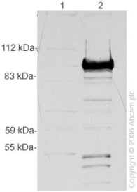
All lanes : Anti-p95/NBS1 antibody (ab23996) at 1/5000 dilution
Lane 1 : Lysate from cells engineered to be NBS1 defective (SV40 transformed, immortal fibroblasts)
Lane 2 : MRC5 cell lysate (SV40 transformed immortal (WT) fibroblasts)
Lysates/proteins at 20 µg per lane.
Performed under reducing conditions.
Predicted band size: 85 kDa
Observed band size: 95 kDa why is the actual band size different from the predicted?
ab23996 detects a band at approximately 95 kDa, the size at which NBS1 migrates, in MRC5 cell lysate. This band is not detected in fibroblasts in which NBS1 is not expressed, indicating that it is specific for the NBS1 protein. -
All lanes : Anti-p95/NBS1 antibody (ab23996) at 1 µg/ml
Lane 1 : HeLa (Human epithelial carcinoma cell line) Whole Cell Lysate
Lane 2 : NIH 3T3 (Mouse embryonic fibroblast cell line) Whole Cell Lysate
Lane 3 : HepG2 (Human hepatocellular liver carcinoma cell line) Whole Cell Lysate
Lane 4 : Jurkat (Human T cell lymphoblast-like cell line) Whole Cell Lysate
Lysates/proteins at 10 µg per lane.
Secondary
All lanes : Goat Anti-Rabbit IgG H&L (HRP) preadsorbed (ab97080) at 1/5000 dilution
Developed using the ECL technique.
Performed under reducing conditions.
Predicted band size: 85 kDa
Observed band size: 95 kDa why is the actual band size different from the predicted?
Additional bands at: 125 kDa, 55 kDa, 65 kDa. We are unsure as to the identity of these extra bands. -
ICC/IF image of ab23996 stained HeLa cells. The cells were 100% methanol fixed (5 min) and then incubated in 1%BSA / 10% normal goat serum / 0.3M glycine in 0.1% PBS-Tween for 1h to permeabilise the cells and block non-specific protein-protein interactions. The cells were then incubated with the antibody (ab23996, 1µg/ml) overnight at +4°C. The secondary antibody (green) was ab96899, DyLight® 488 goat anti-rabbit IgG (H+L) used at a 1/250 dilution for 1h. Alexa Fluor® 594 WGA was used to label plasma membranes (red) at a 1/200 dilution for 1h. DAPI was used to stain the cell nuclei (blue) at a concentration of 1.43µM. This antibody also gave a positive result in 100% methanol fixed (5 min) HepG2 and MCF7 cells at 1µg/ml.
Protocols
Datasheets and documents
References (13)
ab23996 has been referenced in 13 publications.
- Ha Thi HT et al. SMAD7 in keratinocytes promotes skin carcinogenesis by activating ATM-dependent DNA repair and an EGFR-mediated cell proliferation pathway. Carcinogenesis 40:112-120 (2019). PubMed: 30219864
- Ha Thi HT et al. MicroRNA-130a modulates a radiosensitivity of rectal cancer by targeting SOX4. Neoplasia 21:882-892 (2019). PubMed: 31387015
- Velichko AK et al. Hypoosmotic stress induces R loop formation in nucleoli and ATR/ATM-dependent silencing of nucleolar transcription. Nucleic Acids Res 47:6811-6825 (2019). PubMed: 31114877
- Hung PJ et al. MRI Is a DNA Damage Response Adaptor during Classical Non-homologous End Joining. Mol Cell 71:332-342.e8 (2018). PubMed: 30017584
- Bártová E et al. Depletion of A-type lamins and Lap2a reduces 53BP1 accumulation at UV-induced DNA lesions and Lap2a protein is responsible for compactness of irradiated chromatin. J Cell Biochem N/A:N/A (2018). PubMed: 29923310
Images
-
All lanes : Anti-p95/NBS1 antibody (ab23996) at 1/5000 dilution
Lane 1 : Lysate from cells engineered to be NBS1 defective (SV40 transformed, immortal fibroblasts)
Lane 2 : MRC5 cell lysate (SV40 transformed immortal (WT) fibroblasts)
Lysates/proteins at 20 µg per lane.
Performed under reducing conditions.
Predicted band size: 85 kDa
Observed band size: 95 kDa why is the actual band size different from the predicted?
ab23996 detects a band at approximately 95 kDa, the size at which NBS1 migrates, in MRC5 cell lysate. This band is not detected in fibroblasts in which NBS1 is not expressed, indicating that it is specific for the NBS1 protein. -
All lanes : Anti-p95/NBS1 antibody (ab23996) at 1 µg/ml
Lane 1 : HeLa (Human epithelial carcinoma cell line) Whole Cell Lysate
Lane 2 : NIH 3T3 (Mouse embryonic fibroblast cell line) Whole Cell Lysate
Lane 3 : HepG2 (Human hepatocellular liver carcinoma cell line) Whole Cell Lysate
Lane 4 : Jurkat (Human T cell lymphoblast-like cell line) Whole Cell Lysate
Lysates/proteins at 10 µg per lane.
Secondary
All lanes : Goat Anti-Rabbit IgG H&L (HRP) preadsorbed (ab97080) at 1/5000 dilution
Developed using the ECL technique.
Performed under reducing conditions.
Predicted band size: 85 kDa
Observed band size: 95 kDa why is the actual band size different from the predicted?
Additional bands at: 125 kDa, 55 kDa, 65 kDa. We are unsure as to the identity of these extra bands.
-
ICC/IF image of ab23996 stained HeLa cells. The cells were 100% methanol fixed (5 min) and then incubated in 1%BSA / 10% normal goat serum / 0.3M glycine in 0.1% PBS-Tween for 1h to permeabilise the cells and block non-specific protein-protein interactions. The cells were then incubated with the antibody (ab23996, 1µg/ml) overnight at +4°C. The secondary antibody (green) was ab96899, DyLight® 488 goat anti-rabbit IgG (H+L) used at a 1/250 dilution for 1h. Alexa Fluor® 594 WGA was used to label plasma membranes (red) at a 1/200 dilution for 1h. DAPI was used to stain the cell nuclei (blue) at a concentration of 1.43µM. This antibody also gave a positive result in 100% methanol fixed (5 min) HepG2 and MCF7 cells at 1µg/ml.


