Anti-p38 (phospho T180 + Y182) antibody (ab4822)
Key features and details
- Rabbit polyclonal to p38 (phospho T180 + Y182)
- Suitable for: IHC-P, ICC/IF, WB
- Reacts with: Rat, Human
- Isotype: IgG
Overview
-
Product name
Anti-p38 (phospho T180 + Y182) antibody
See all p38 primary antibodies -
Description
Rabbit polyclonal to p38 (phospho T180 + Y182) -
Host species
Rabbit -
Tested Applications & Species
See all applications and species dataApplication Species ICC/IF HumanIHC-P RatHumanWB Human -
Immunogen
-
Positive control
- HEK 293 cells +/- UV irradiation treatment and PC12 cells +/- Sorbitol.
-
General notes
The Life Science industry has been in the grips of a reproducibility crisis for a number of years. Abcam is leading the way in addressing the problem with our range of recombinant monoclonal antibodies and knockout edited cell lines for gold-standard validation.
One factor contributing to the crisis is the use of antibodies that are not suitable. This can lead to misleading results and the use of incorrect data informing project assumptions and direction. To help address this challenge, we have introduced an application and species grid on our primary antibody datasheets to make it easy to simplify identification of the right antibody for your needs.
Learn more here.
Properties
-
Form
Liquid -
Storage instructions
Shipped at 4°C. Store at +4°C short term (1-2 weeks). Upon delivery aliquot. Store at -20°C. Avoid freeze / thaw cycle. -
Storage buffer
pH: 7.30
Preservative: 0.05% Sodium azide
Constituents: PBS, 50% Glycerol, 0.1% BSA
BSA is IgG and protease free. PBS without Mg2+ and Ca2+. -
 Concentration information loading...
 -
Purity
Protein A purified -
Purification notes
Purified from rabbit serum by sequential epitope-specific chromatography. The antibody has been negatively preadsorbed using i) non-phosphopeptide corresponding to the site of phosphorylation to remove antibody that is reactive with non-phosphorylated p38, and ii) a JNK-derived peptide that is phosphorylated at threonine 183 and tyrosine 185. The final product is generated by affinity chromatography using a p38-derived peptide that is phosphorylated at threonine 180 and tyrosine 182. -
Clonality
Polyclonal -
Isotype
IgG -
Research areas
Images
-
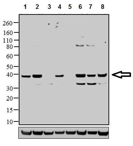
All lanes : Anti-p38 (phospho T180 + Y182) antibody (ab4822) at 1/1000 dilution
Lane 1 : HeLa (human epithelial cell line from cervix adenocarcinoma) cell lysate
Lane 2 : HeLa (human epithelial cell line from cervix adenocarcinoma) exposed for 40 min with UV, cell lysate
Lane 3 : A431 (human epidermoid carcinoma cell line) cell lysate
Lane 4 : A431 (human epidermoid carcinoma cell line) exposed for 40 min with UV, cell lysate
Lane 5 : COLO 205 (human colon adenocarcinoma cell line) cell lysate
Lane 6 : COLO 205 (human colon adenocarcinoma cell line) exposed for 40 min with UV, cell lysate
Lane 7 : A549 (human lung carcinoma cell line) cell lysate
Lane 8 : A549 (human lung carcinoma cell line) exposed for 40 min with UV, cell lysate
Lysates/proteins at 20 µg per lane.
Secondary
All lanes : Goat anti-Rabbit IgG HRP at 1/5000 dilution
Developed using the ECL technique.
Predicted band size: 38 kDa
-
 Immunohistochemistry (Formalin/PFA-fixed paraffin-embedded sections) - Anti-p38 (phospho T180 + Y182) antibody (ab4822)
Paraffin-embedded human brain tissue stained for p38 (phospho T180 + Y182) using ab4822 (right panel) at 1/100 dilution in immunohistochemical analysis followed by HRP-conjugated secondary antibody and DAB staining. Negative control (left panel) staining without primary antibody.
-
 Immunohistochemistry (Formalin/PFA-fixed paraffin-embedded sections) - Anti-p38 (phospho T180 + Y182) antibody (ab4822)
Paraffin-embedded human heart tissue stained for p38 (phospho T180 + Y182) using ab4822 (right panel) at 1/20 dilution in immunohistochemical analysis followed by HRP-conjugated secondary antibody and DAB staining. Negative control (left panel) staining without primary antibody.
-
 Immunohistochemistry (Formalin/PFA-fixed paraffin-embedded sections) - Anti-p38 (phospho T180 + Y182) antibody (ab4822)
Paraffin-embedded rat heart tissue stained for p38 (phospho T180 + Y182) using ab4822 (right panel) at 1/20 dilution in immunohistochemical analysis followed by HRP-conjugated secondary antibody and DAB staining. Negative control (left panel) staining without primary antibody.
-
4% PFA-fixed, Triton X-100 permeabilized SH-SY5Y (human neuroblastoma cell line from bone marrow) cells labeling p38 (phospho T180 + Y182) (Panel A: green) using ab4822 at 1 µg/mL in ICC/IF. Secondary antibody: Alexa Flour® 488 Goat Anti-Rabbit IgG at 1/400 dilution. Nuclei (Panel b: blue) were stained with SlowFade® Gold Antifade Mountant with DAPI. F-actin (Panel c: red) was stained with Alexa Fluor® 594 Phalloidin. Panel d is a merged image showing nuclear localization. Panel e is a no primary antibody control.
-
Peptide Competition: Extracts prepared from HEK-293 (human epithelial cell line from embryonic kidney) cells treated with UV irradiation were resolved on a 10% Tris-glycine gel and transferred to nitrocellulose. Membranes were blocked with a 5% BSA-TBST buffer overnight at 4oC, then were incubated with 0.50 µg/mL ab4822 for two hours at room temperature in a 3% BSA-TBST buffer, following its prior incubation with: the peptide immunogen (1), a generic phosphothreonine containing peptide (2), a generic phosphotyrosine-containing peptide (3), the non-phosphorylated peptide corresponding to the phosphopeptide (4), no peptide (5), the phosphorylated peptide derived from the corresponding region of JNK 1 & 2 (6), and, the phosphorylated peptide derived from the corresponding region of ERK 1 & 2 (7). After washing, membranes were incubated with goat F(ab')2 antirabbit IgG alkaline phosphatase and the signal was detected using the Tropix WesternStar method. The data show that only the phosphopeptide


