Anti-XIAP antibody (ab21278)
")
Key features and details
- Rabbit polyclonal to XIAP
- Suitable for: IHC-P, ICC/IF, WB
- Reacts with: Mouse, Human
- Isotype: IgG
Overview
-
Product name
Anti-XIAP antibody
See all XIAP primary antibodies -
Description
Rabbit polyclonal to XIAP -
Host species
Rabbit -
Tested Applications & Species
See all applications and species dataApplication Species ICC/IF HumanIHC-P MouseHumanWB Human
-
Immunogen
Synthetic peptide corresponding to 13 amino acids at the C-terminus of human XIAP.
-
Positive control
- Human kidney cell lysate.
-
General notes
The Life Science industry has been in the grips of a reproducibility crisis for a number of years. Abcam is leading the way in addressing the problem with our range of recombinant monoclonal antibodies and knockout edited cell lines for gold-standard validation.
One factor contributing to the crisis is the use of antibodies that are not suitable. This can lead to misleading results and the use of incorrect data informing project assumptions and direction. To help address this challenge, we have introduced an application and species grid on our primary antibody datasheets to make it easy to simplify identification of the right antibody for your needs.
Learn more here.
Properties
-
Form
Liquid -
Storage instructions
Shipped at 4°C. Store at +4°C. -
Storage buffer
pH: 7.2
Preservative: 0.02% Sodium azide
Constituent: PBS -
 Concentration information loading...
 -
Purification notes
Purified by ion exchange chromatography. -
Clonality
Polyclonal -
Isotype
IgG -
Research areas
Images
-
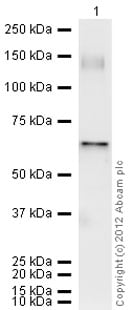
Western blot - Anti-XIAP antibody (ab21278)Lane 1 : Anti-XIAP antibody (ab21278) at 0.5 µg/ml
Lane 2 : Anti-XIAP antibody (ab21278) at 1 µg/ml
Lane 3 : Anti-XIAP antibody (ab21278) at 2 µg/ml
All lanes : Human kidney lysate
Predicted band size: 55 kDaWestern blot analysis of XIAP in human kidney cell lysate using ab21278 at a concentration of 0.5
µ g/ml (lane A), 1µ g/ml (lane B) and 2µ g/ml (lane C). -
 Immunohistochemistry (Formalin/PFA-fixed paraffin-embedded sections) - Anti-XIAP antibody (ab21278)Immunohistochemistry (Formalin/PFA-fixed paraffin-embedded sections) of mouse kidney tissue staining XIAP with ab21278 at 20µg/ml
 -
 Immunohistochemistry (Formalin/PFA-fixed paraffin-embedded sections) - Anti-XIAP antibody (ab21278)Immunohistochemistry (Formalin-fixed paraffin embedded sections) of mouse kidney tissue labeling XIAP with Anti-XIAP antibody (ab21278) at 5μg/ml.
 -
 Immunohistochemistry (Formalin/PFA-fixed paraffin-embedded sections) - Anti-XIAP antibody (ab21278)
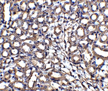
ab21278 at 2µg/ml staining XIAP in human kidney tissue by IHC
-
 Immunocytochemistry/ Immunofluorescence - Anti-XIAP antibody (ab21278)ICC/IF image of ab21278 stained HeLa cells. The cells were 4% formaldehyde fixed (10 min) and then incubated in 1%BSA / 10% normal goat serum / 0.3M glycine in 0.1% PBS-Tween for 1h to permeabilise the cells and block non-specific protein-protein interactions. The cells were then incubated with the antibody (ab21278, 5µg/ml) overnight at +4°C. The secondary antibody (green) was Alexa Fluor® 488 goat anti-rabbit IgG (H+L) used at a 1/1000 dilution for 1h. Alexa Fluor® 594 WGA was used to label plasma membranes (red) at a 1/200 dilution for 1h. DAPI was used to stain the cell nuclei (blue) at a concentration of 1.43µM.
-
 Western blot - Anti-XIAP antibody (ab21278)


