Anti-LDL Receptor antibody (ab30532)
")
Key features and details
- Rabbit polyclonal to LDL Receptor
- Suitable for: IHC-P, ICC/IF, WB
- Reacts with: Mouse, Rat, Human
- Isotype: IgG
Overview
-
Product name
Anti-LDL Receptor antibody
See all LDL Receptor primary antibodies -
Description
Rabbit polyclonal to LDL Receptor -
Host species
Rabbit -
Tested Applications & Species
See all applications and species dataApplication Species ICC/IF HumanIHC-P MouseHumanWB MouseHuman
-
Immunogen
Synthetic peptide:
SVADTKGVKRRTL
, corresponding to amino acids 499-511 of Mouse LDL Receptor -
General notes
The Life Science industry has been in the grips of a reproducibility crisis for a number of years. Abcam is leading the way in addressing the problem with our range of recombinant monoclonal antibodies and knockout edited cell lines for gold-standard validation.
One factor contributing to the crisis is the use of antibodies that are not suitable. This can lead to misleading results and the use of incorrect data informing project assumptions and direction. To help address this challenge, we have introduced an application and species grid on our primary antibody datasheets to make it easy to simplify identification of the right antibody for your needs.
Learn more here.


Properties
-
Form
Liquid -
Storage instructions
Shipped at 4°C. Upon delivery aliquot and store at -20°C. Avoid freeze / thaw cycles. -
Storage buffer
pH: 7.40
Preservative: 0.02% Sodium azide
Constituents: Tris buffered saline, 40% Glycerol, 0.1% BSA -
 Concentration information loading...
 -
Purity
Immunogen affinity purified -
Clonality
Polyclonal -
Isotype
IgG -
Research areas
- Metabolism
- Pathways and Processes
- Metabolic signaling pathways
- Lipid and lipoprotein metabolism
- Lipid metabolism
- Metabolism
- Pathways and Processes
- Metabolic signaling pathways
- Lipid and lipoprotein metabolism
- Cholesterol Metabolism
Images
-
Western blot - Anti-LDL Receptor antibody (ab30532)All lanes : Anti-LDL Receptor antibody (ab30532)
Lane 1 : RAW 264.7 Cells at 25 µg
Lane 2 : RAW 264.7 Cells at 50 µg
Lane 3 : Rat Liver Supernatant at 50 µg
Predicted band size: 95 kDa
-
 Immunohistochemistry (Formalin/PFA-fixed paraffin-embedded sections) - Anti-LDL Receptor antibody (ab30532)
Immunohistochemistry analysis of formalin-fixed, paraffin-embedded (FFPE) mouse lung tissue after heat induced antigen retrieval in pH 6.0 citrate buffer. After incubation with LDL receptor polyclonal antibody, ab30532, at a 1:100 dilution, slides were incubated with biotinylated secondary antibody, followed by alkaline phosphatase-streptavidin and chromogen (DAB).
-
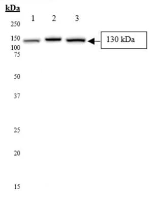 Western blot - Anti-LDL Receptor antibody (ab30532)All lanes : Anti-LDL Receptor antibody (ab30532)
Lane 1 : HeLa Cell Lysate
Lane 2 : HepG2 Cell Lysate
Lane 3 : Huh7 Cell Lysate
Lysates/proteins at 50 µg per lane.
Secondary
All lanes : Mouse monoclonal [6F12] to Cytomegalovirus (ab6500) (Anti-Rabbit IgG (HRP) conjugate)
Predicted band size: 95 kDa
-
 Immunohistochemistry (Formalin/PFA-fixed paraffin-embedded sections) - Anti-LDL Receptor antibody (ab30532)
ab30532 staining LDL receptor in human lung parenchyma.
Left panel: with primary antibody at 1/400. Right panel: isotype control.
Sections were stained using an automated system (DAKO Autostainer Plus), at room temperature: sections were rehydrated and antigen retrieved with the Dako 3 in 1 AR buffers citrate pH6.1. Slides were peroxidase blocked in 3% H2O2 in methanol for 10 mins. They were then blocked with Dako Protein block for 10 minutes (containing casein 0.25% in PBS) then incubated with primary antibody for 20 min and detected with Dako envision flex amplification kit for 30 minutes. Colorimetric detection was completed with Diaminobenzidine for 5 minutes. Slides were counterstained with Haematoxylin and coverslipped under DePeX. Please note that for manual staining we recommend to optimize the primary antibody concentration and incubation time (overnight incubation), and amplification may be required. -
 Immunocytochemistry/ Immunofluorescence - Anti-LDL Receptor antibody (ab30532)
ICC/IF image of ab30532 stained HepG2 cells. The cells were 4% PFA fixed (10 min) and then incubated in 1%BSA / 10% normal goat serum / 0.3M glycine in 0.1% PBS-Tween for 1h to permeabilise the cells and block non-specific protein-protein interactions. The cells were then incubated with the antibody (ab30532, 1/400) overnight at +4°C. The secondary antibody (green) was Alexa Fluor® 488 goat anti-rabbit IgG (H+L) used at a 1/1000 dilution for 1h. Alexa Fluor® 594 WGA was used to label plasma membranes (red) at a 1/200 dilution for 1h. DAPI was used to stain the cell nuclei (blue) at a concentration of 1.43µM.


