Anti-Superoxide Dismutase 1 antibody (ab45777)
Key features and details
- Rabbit polyclonal to Superoxide Dismutase 1
- Suitable for: ICC/IF, WB
- Reacts with: Human
- Isotype: IgG
Overview
-
Product name
Anti-Superoxide Dismutase 1 antibody
See all Superoxide Dismutase 1 primary antibodies -
Description
Rabbit polyclonal to Superoxide Dismutase 1 -
Host species
Rabbit -
Tested Applications & Species
See all applications and species dataApplication Species ICC/IF HumanWB Human -
Immunogen
Synthetic peptide conjugated to KLH derived from within residues 1 - 100 of Human Superoxide Dismutase 1.
Read Abcam's proprietary immunogen policy (Peptides available as ab46993).
Properties
-
Form
Liquid -
Storage instructions
Shipped at 4°C. Store at +4°C short term (1-2 weeks). Upon delivery aliquot. Store at -20°C or -80°C. Avoid freeze / thaw cycle. -
Storage buffer
pH: 7.40
Preservative: 0.02% Sodium azide
Constituent: PBS
Batches of this product that have a concentration Concentration information loading...
Purity
Immunogen affinity purifiedClonality
PolyclonalIsotype
IgGResearch areas
Associated products
-
Compatible Secondaries
-
Isotype control
-
Positive Controls
-
Recombinant Protein
Applications
The Abpromise guarantee
Our Abpromise guarantee covers the use of ab45777 in the following tested applications.
The application notes include recommended starting dilutions; optimal dilutions/concentrations should be determined by the end user.
GuaranteedTested applications are guaranteed to work and covered by our Abpromise guarantee.
PredictedPredicted to work for this combination of applications and species but not guaranteed.
IncompatibleDoes not work for this combination of applications and species.
Application Species ICC/IF HumanWB HumanApplication Abreviews Notes ICC/IF Use a concentration of 1 µg/ml.WB (1) Use a concentration of 0.5 µg/ml. Detects a band of approximately 18 kDa (predicted molecular weight: 16 kDa).Notes ICC/IF
Use a concentration of 1 µg/ml.WB
Use a concentration of 0.5 µg/ml. Detects a band of approximately 18 kDa (predicted molecular weight: 16 kDa).Target
-
Function
Destroys radicals which are normally produced within the cells and which are toxic to biological systems. -
Involvement in disease
Defects in SOD1 are the cause of amyotrophic lateral sclerosis type 1 (ALS1) [MIM:105400]. ALS1 is a familial form of amyotrophic lateral sclerosis, a neurodegenerative disorder affecting upper and lower motor neurons and resulting in fatal paralysis. Sensory abnormalities are absent. Death usually occurs within 2 to 5 years. The etiology of amyotrophic lateral sclerosis is likely to be multifactorial, involving both genetic and environmental factors. The disease is inherited in 5-10% of cases leading to familial forms. -
Sequence similarities
Belongs to the Cu-Zn superoxide dismutase family. -
Post-translational
modificationsUnlike wild-type protein, the pathogenic variants ALS1 Arg-38, Arg-47, Arg-86 and Ala-94 are polyubiquitinated by RNF19A leading to their proteasomal degradation. The pathogenic variants ALS1 Arg-86 and Ala-94 are ubiquitinated by MARCH5 leading to their proteasomal degradation.
The ditryptophan cross-link at Trp-33 is reponsible for the non-disulfide-linked homodimerization. Such modification might only occur in extreme conditions and additional experimental evidence is required. -
Cellular localization
Cytoplasm. The pathogenic variants ALS1 Arg-86 and Ala-94 gradually aggregates and accumulates in mitochondria. - Information by UniProt
-
Database links
- Entrez Gene: 6647 Human
- Omim: 147450 Human
- SwissProt: P00441 Human
- Unigene: 443914 Human
-
Alternative names
- ALS antibody
- ALS1 antibody
- Amyotrophic lateral sclerosis 1 adult antibody
see all
Images
-
ICC/IF image of ab45777 stained human HeLa cells. The cells were methanol fixed (5 min), permabilised in TBS-T (20 min) and incubated with the antibody (ab45777, 1µg/ml) for 1h at room temperature. 1%BSA / 10% normal serum / 0.3M glycine was used to quench autofluorescence and block non-specific protein-protein interactions. The secondary antibody (green) was Alexa Fluor® 488 goat anti-rabbit IgG (H+L) used at a 1/1000 dilution for 1h. Alexa Fluor® 594 WGA was used to label plasma membranes (red). DAPI was used to stain the cell nuclei (blue).
-
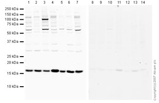
 Western blot - Anti-Superoxide Dismutase 1 antibody (ab45777)This image is courtesy of an Abreview submitted by Dr Lay Harn GamAll lanes : Anti-Superoxide Dismutase 1 antibody (ab45777) at 1/1000 dilution
All lanes : Whole tissue lysate prepared from human breast cancer tissue
Lysates/proteins at 8 µg per lane.
Secondary
All lanes : HRP conjugated goat polyclonal to rabbit IgG at 1/1000 dilution
Performed under reducing conditions.
Predicted band size: 16 kDa
Exposure time: 15 minutes
Protocols
Datasheets and documents
References (0)
ab45777 has not yet been referenced specifically in any publications.
Images
-
-
ICC/IF image of ab45777 stained human HeLa cells. The cells were methanol fixed (5 min), permabilised in TBS-T (20 min) and incubated with the antibody (ab45777, 1µg/ml) for 1h at room temperature. 1%BSA / 10% normal serum / 0.3M glycine was used to quench autofluorescence and block non-specific protein-protein interactions. The secondary antibody (green) was Alexa Fluor® 488 goat anti-rabbit IgG (H+L) used at a 1/1000 dilution for 1h. Alexa Fluor® 594 WGA was used to label plasma membranes (red). DAPI was used to stain the cell nuclei (blue).
-
 Western blot - Anti-Superoxide Dismutase 1 antibody (ab45777) This image is courtesy of an Abreview submitted by Dr Lay Harn GamAll lanes : Anti-Superoxide Dismutase 1 antibody (ab45777) at 1/1000 dilution
All lanes : Whole tissue lysate prepared from human breast cancer tissue
Lysates/proteins at 8 µg per lane.
Secondary
All lanes : HRP conjugated goat polyclonal to rabbit IgG at 1/1000 dilution
Performed under reducing conditions.
Predicted band size: 16 kDa
Exposure time: 15 minutes


