Anti-Histone H2A.Z antibody - ChIP Grade (ab4174)
Key features and details
- Rabbit polyclonal to Histone H2A.Z - ChIP Grade
- Suitable for: ICC/IF, ChIP, WB
- Reacts with: Mouse, Rat, Cow, Human
- Isotype: IgG
Overview
-
Product name
Anti-Histone H2A.Z antibody - ChIP Grade
See all Histone H2A.Z primary antibodies -
Description
Rabbit polyclonal to Histone H2A.Z - ChIP Grade -
Host species
Rabbit -
Tested Applications & Species
See all applications and species dataApplication Species ChIP HumanICC/IF MouseHumanWB Cow -
Immunogen
Synthetic peptide corresponding to Human Histone H2A.Z aa 100 to the C-terminus conjugated to keyhole limpet haemocyanin.
(Peptide available asab11681)
Images
-
 ChIP - Anti-Histone H2A.Z antibody - ChIP Grade (ab4174) Gjidoda et al PLoS One. 2014 Apr 4;9(4):e93971. doi: 10.1371/journal.pone.0093971. eCollection 2014. Fig 2. Reproduced under the Creative Commons license http://creativecommons.org/licenses/by/4.0/
Changes in nucleosome occupancy upon LPS induction at a putative distal enhancer and promoter of IL1A, kinetics of nucleosome removal, and changes in histone modifications.
Chromation from mouse bone marrow derived macrophages. ChIP experiments with antibodies against H3 (dark blue bars), H2A.Z (light blue, (ab4174 at 4µg), H3K4me1 (green, ab8895 at 1 µg), H3K4me3 (yellow, ab8580 at 1 µg) and H3K27ac (red, ab4729 at 1 µg). For these experiments cross-linked chromatin was lightly digested with MNase before incubation with the respective antibodies to increase resolution of the ChIP signal and the data was normalized to a region in the ORF of RPL4. Changes upon LPS induction in histone binding and histone modifications at the enhancers and promoters of IL12B and IL1A as well as at a control region in the GAPDH pseudo gene are shown as fold over levels found before induction. For H3K27ac the changes 1.5 h after LPS induction, and for all other histone variants and modifications the changes after 3 h of induction are shown. The error bars show the SEM of at least 3 independent experiments. Statistical significance of the changes in H3K4me3 and H3K27ac upon LPS induction compared to levels found prior to induction determined by Student's T-tests is indicated (*P
-
Chromatin was prepared from HeLa (Human epithelial cell line from cervix adenocarcinoma) cells according to the Abcam X-ChIP protocol.
Cells were fixed with formaldehyde for 10 min. The ChIP was performed with 25 µg of chromatin, 2 µg of ab4174 (blue), and 20 µl of Protein A/G sepharose beads. No antibody was added to the beads control (yellow). The immunoprecipitated DNA was quantified by real time PCR (Taqman approach for active and inactive loci, Sybr green approach for heterochromatic loci). Primers and probes are located in the first kb of the transcribed region.
-
Lanes 1-2 : Anti-Histone H2A.Z antibody - ChIP Grade (ab4174) at 1/500 dilution
Lanes 3-4 : Anti-Histone H2A.Z antibody - ChIP Grade (ab4174) at 1/1000 dilution
Lanes 1 & 3 : Calf thymus histone lysate
Lanes 2 & 4 : Calf thymus histone lysate withHuman Histone H2A.Z peptide (ab11681) at 1 µg/ml
Secondary
All lanes : Goat Anti-Rabbit IgG H&L (HRP) (ab6721) at 1/5000 dilution
Performed under reducing conditions.
Predicted band size: 13.4 kDa -
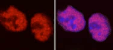
Staining of Histone H2A.Z in mouse embryonic cells. The fixation is 2% PFA, and permeabilization is PBS 0.5% triton BSA. The dilution used was 1 in 100 (but it could be used at 1/200 to 1/300).
Red = H2A.Z
Blue = toto3 for the DNA -
All lanes : Anti-Histone H2A.Z antibody - ChIP Grade (ab4174) at 1 µg/ml
Lane 1 : Calf Thymus Histone Preparation Nuclear Lysate at 0.5 µg
Lane 2 : HeLa (Human epithelial carcinoma cell line) Whole Cell Lysate at 10 µg
Lane 3 : NIH/3T3 (Mouse embryonic fibroblast cell line) Whole Cell Lysate at 10 µg
Lane 4 : PC-12 (Rat adrenal pheochromocytoma cell line) Whole Cell Lysate at 10 µg
Secondary
All lanes : Goat polyclonal to Rabbit IgG - H&L - Pre-Adsorbed (HRP) (ab65484) at 1/3000 dilution
Developed using the ECL technique.
Performed under reducing conditions.
Predicted band size: 13.4 kDa
Exposure time: 3 minutes
-
ICC/IF image of ab4174 stained HepG2 (Human liver hepatocellular carcinoma cell line) cells.
The cells were fixed with 100% methanol (5 min) and then incubated in 1%BSA / 10% normal goat serum / 0.3M glycine in 0.1% PBS-Tween for 1h to permeabilize the cells and block non-specific protein-protein interactions. The cells were then incubated with the antibody (ab4174, 5 µg/ml) overnight at +4°C. The secondary antibody (green) was DyLight® 488 goat anti-rabbit IgG - H&L, pre-adsorbed (ab96899) used at a 1/250 dilution for 1h. Alexa Fluor® 594 WGA was used to label plasma membranes (red) at a 1/200 dilution for 1h. DAPI was used to stain the cell nuclei (blue) at a concentration of 1.43µM.
-
 Western blot - Anti-Histone H2A.Z antibody - ChIP Grade (ab4174) This image is courtesy of an Abreview submitted by Ragnhild EskelandAll lanes : Anti-Histone H2A.Z antibody - ChIP Grade (ab4174) at 1/1000 dilution
Lane 1 : Native recombinant octamers K562 (Human chronic myelogenous leukemia cell line from bone marrow) cells at 3 µg
Lane 2 : Native recombinant octamers K562 cells at 1.5 µg
Lane 3 : Recombinant Human octamers containing H2A at 1 µg
Lane 4 : Recombinant Human octamers containing H2A at 0.5 µg
Lane 5 : Recombinant Human octamers containing H2A.Z.2.1 at 0.5 µg
Lane 6 : Recombinant Human octamers containing H2A.Z.1 at 0.5 µg
Secondary
All lanes : HRP-conjugated donkey anti-rabbit IgG polyclonal at 1/10000 dilution
Developed using the ECL technique.
Performed under reducing conditions.
Predicted band size: 13.4 kDa
Exposure time: 5 minutesBlocked with 3% BSA for 1 hour at 20°C.
Primary incubation in TBS tween + 3% BSA at 20°C for 1 hour.


