Anti-Histone H1.2 antibody (ab17677)
")
Key features and details
- Rabbit polyclonal to Histone H1.2
- Suitable for: ICC/IF, IP, WB
- Reacts with: Human, Recombinant fragment
- Isotype: IgG
Overview
-
Product name
Anti-Histone H1.2 antibody
See all Histone H1.2 primary antibodies -
Description
Rabbit polyclonal to Histone H1.2 -
Host species
Rabbit -
Tested applications
Suitable for: ICC/IF, IP, WBmore details -
Species reactivity
Reacts with: Human, Recombinant fragment -
Immunogen
-
Positive control
- This antibody gave a positive signal in Human H1.2 Recombinant Protein. ICC/IF: HeLa cell line
Properties
-
Form
Liquid -
Storage instructions
Shipped at 4°C. Store at +4°C short term (1-2 weeks). Upon delivery aliquot. Store at -20°C or -80°C. Avoid freeze / thaw cycle. -
Storage buffer
pH: 7.40
Preservative: 0.02% Sodium azide
Constituent: PBS
Batches of this product that have a concentration Concentration information loading...
Purity
Immunogen affinity purifiedClonality
PolyclonalIsotype
IgGResearch areas
Associated products
-
Compatible Secondaries
-
Immunizing Peptide (Blocking)
-
Isotype control
-
Recombinant Protein
Applications
Our Abpromise guarantee covers the use of ab17677 in the following tested applications.
The application notes include recommended starting dilutions; optimal dilutions/concentrations should be determined by the end user.
Application Abreviews Notes ICC/IF Use a concentration of 1 µg/ml. IP Use a concentration of 5 µg/ml. WB 1/1000. Detects a band of approximately 29 kDa (predicted molecular weight: 29 kDa). Target
-
Function
Histones H1 are necessary for the condensation of nucleosome chains into higher order structures. -
Sequence similarities
Belongs to the histone H1/H5 family.
Contains 1 H15 (linker histone H1/H5 globular) domain. -
Cellular localization
Nucleus. Chromosome. - Information by UniProt
-
Database links
- Entrez Gene: 3006 Human
- Omim: 142710 Human
- SwissProt: P16403 Human
- Unigene: 7644 Human
-
Alternative names
- H1 histone family member 2 antibody
- H1.a antibody
- H12_HUMAN antibody
see all
Images
-
Western blot - Anti-Histone H1.2 antibody (ab17677)This image is courtesy of Dr Albert Jordan and Monica Sancho, Center for Genomic Regulation (CRG). Recombinant H1 isoforms courtesy of Dr Nicole Happel.All lanes : Anti-Histone H1.2 antibody (ab17677) at 1/1000 dilution
Lane 1 : Recombinant histone H1
Lane 2 : Recombinant histone H1.1
Lane 3 : Recombinant histone H1.2
Lane 4 : Recombinant histone H1.3
Lane 5 : Recombinant histone H1.4
Lane 6 : Recombinant histone H1.5
Predicted band size: 29 kDa
Observed band size: 29 kDa -
 Immunoprecipitation - Anti-Histone H1.2 antibody (ab17677)
Histone H1.2 was immunoprecipitated using 0.5mg Hela whole cell extract, 5µg of Rabbit polyclonal to Histone H1.2 and 50µl of protein G magnetic beads (+). No antibody was added to the control (-).
The antibody was incubated under agitation with Protein G beads for 10min, Hela whole cell extract lysate diluted in RIPA buffer was added to each sample and incubated for a further 10min under agitation.
Proteins were eluted by addition of 40µl SDS loading buffer and incubated for 10min at 70°C; 10µl of each sample was separated on a SDS PAGE gel, transferred to a nitrocellulose membrane, blocked with 5% BSA and probed with ab17677.
Secondary: Mouse monoclonal [SB62a] Secondary Antibody to Rabbit IgG light chain (HRP) (ab99697).
Band: 35kDa; Histone H1.2
-
 Immunocytochemistry/ Immunofluorescence - Anti-Histone H1.2 antibody (ab17677)
ab17677 staining Histone H1.2 in HeLa cells. The cells were fixed with 100% methanol (5 min) at room temperature, permeabilized with 0.1% Triton X-100 for 5 minutes and then blocked with 1% BSA/10% normal goat serum/0.3M glycine in 0.1% PBS-Tritonn for 1h. The cells were then incubated with the antibody ab16766 at 0.1µg/ml and ab7291 (Mouse monoclonal to alpha Tubulin - Loading Control) used at a 1/1000 dilution overnight at +4°C. The secondary antibodies were ab150081, Goat Anti-Rabbit IgG H&L (Alexa Fluor® 488) preadsorbed, (pseudo-colored green) and ab150120, Goat polyclonal Secondary Antibody to Mouse IgG - H&L (Alexa Fluor® 594) preadsorbed, (colored red), both used at a 1/1000 dilution for 1 hour at room temperature. DAPI was used to stain the cell nuclei (colored blue) at a concentration of 1.43 µM for 1hour at room temperature.
-
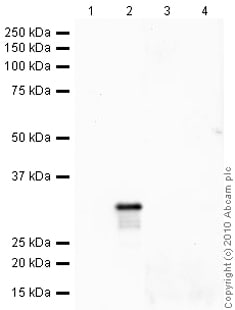 Western blot - Anti-Histone H1.2 antibody (ab17677)All lanes : Anti-Histone H1.2 antibody (ab17677) at 1 µg/ml
Lane 1 : Histone H1 Recombinant Protein
Lane 2 : Histone H1.2 (Human) - Recombinant Protein
Lane 3 : Histone H1 Recombinant Protein withHuman Histone H1.2 peptide (ab18500) at 1 µg/ml
Lane 4 : Histone H1.2 (Human) - Recombinant Protein withHuman Histone H1.2 peptide (ab18500) at 1 µg/ml
Lysates/proteins at 0.1 µg per lane.
Secondary
All lanes : Goat polyclonal to Rabbit IgG - H&L - Pre-Adsorbed (HRP) at 1/3000 dilution
Developed using the ECL technique.
Performed under reducing conditions.
Predicted band size: 29 kDa
Observed band size: 29 kDa
Exposure time: 12 minutes
Protocols
References (27)
ab17677 has been referenced in 27 publications.
- Bernstein DI et al. Genetic variants with gene regulatory effects are associated with diisocyanate-induced asthma. J Allergy Clin Immunol 142:959-969 (2018). PubMed: 29969634
- Li Z et al. Destabilization of linker histone H1.2 is essential for ATM activation and DNA damage repair. Cell Res 28:756-770 (2018). PubMed: 29844578
- Liu Q et al. 12-HETE facilitates cell survival by activating the integrin-linked kinase/NF-?B pathway in ovarian cancer. Cancer Manag Res 10:5825-5838 (2018). PubMed: 30510451
- Clouaire T et al. Comprehensive Mapping of Histone Modifications at DNA Double-Strand Breaks Deciphers Repair Pathway Chromatin Signatures. Mol Cell 72:250-262.e6 (2018). PubMed: 30270107
- Chen J et al. The linker histone H1.2 is a novel component of the nucleolar organizer regions. J Biol Chem 293:2358-2369 (2018). PubMed: 29301938
Images
-
Western blot - Anti-Histone H1.2 antibody (ab17677) This image is courtesy of Dr Albert Jordan and Monica Sancho, Center for Genomic Regulation (CRG). Recombinant H1 isoforms courtesy of Dr Nicole Happel.All lanes : Anti-Histone H1.2 antibody (ab17677) at 1/1000 dilution
Lane 1 : Recombinant histone H1
Lane 2 : Recombinant histone H1.1
Lane 3 : Recombinant histone H1.2
Lane 4 : Recombinant histone H1.3
Lane 5 : Recombinant histone H1.4
Lane 6 : Recombinant histone H1.5
Predicted band size: 29 kDa
Observed band size: 29 kDa
-
Immunoprecipitation - Anti-Histone H1.2 antibody (ab17677)
Histone H1.2 was immunoprecipitated using 0.5mg Hela whole cell extract, 5µg of Rabbit polyclonal to Histone H1.2 and 50µl of protein G magnetic beads (+). No antibody was added to the control (-).
The antibody was incubated under agitation with Protein G beads for 10min, Hela whole cell extract lysate diluted in RIPA buffer was added to each sample and incubated for a further 10min under agitation.
Proteins were eluted by addition of 40µl SDS loading buffer and incubated for 10min at 70°C; 10µl of each sample was separated on a SDS PAGE gel, transferred to a nitrocellulose membrane, blocked with 5% BSA and probed with ab17677.
Secondary: Mouse monoclonal [SB62a] Secondary Antibody to Rabbit IgG light chain (HRP) (ab99697).
Band: 35kDa; Histone H1.2
-
Immunocytochemistry/ Immunofluorescence - Anti-Histone H1.2 antibody (ab17677)
ab17677 staining Histone H1.2 in HeLa cells. The cells were fixed with 100% methanol (5 min) at room temperature, permeabilized with 0.1% Triton X-100 for 5 minutes and then blocked with 1% BSA/10% normal goat serum/0.3M glycine in 0.1% PBS-Tritonn for 1h. The cells were then incubated with the antibody ab16766 at 0.1µg/ml and ab7291 (Mouse monoclonal to alpha Tubulin - Loading Control) used at a 1/1000 dilution overnight at +4°C. The secondary antibodies were ab150081, Goat Anti-Rabbit IgG H&L (Alexa Fluor® 488) preadsorbed, (pseudo-colored green) and ab150120, Goat polyclonal Secondary Antibody to Mouse IgG - H&L (Alexa Fluor® 594) preadsorbed, (colored red), both used at a 1/1000 dilution for 1 hour at room temperature. DAPI was used to stain the cell nuclei (colored blue) at a concentration of 1.43 µM for 1hour at room temperature.
-
Western blot - Anti-Histone H1.2 antibody (ab17677)All lanes : Anti-Histone H1.2 antibody (ab17677) at 1 µg/ml
Lane 1 : Histone H1 Recombinant Protein
Lane 2 : Histone H1.2 (Human) - Recombinant Protein
Lane 3 : Histone H1 Recombinant Protein withHuman Histone H1.2 peptide (ab18500) at 1 µg/ml
Lane 4 : Histone H1.2 (Human) - Recombinant Protein withHuman Histone H1.2 peptide (ab18500) at 1 µg/ml
Lysates/proteins at 0.1 µg per lane.
Secondary
All lanes : Goat polyclonal to Rabbit IgG - H&L - Pre-Adsorbed (HRP) at 1/3000 dilution
Developed using the ECL technique.
Performed under reducing conditions.
Predicted band size: 29 kDa
Observed band size: 29 kDa
Exposure time: 12 minutes


