Anti-ERK1 + ERK2 antibody (ab17942)
Key features and details
- Rabbit polyclonal to ERK1 + ERK2
- Suitable for: ICC, IHC-P, WB
- Reacts with: Mouse, Rat, Human
- Isotype: IgG
Overview
-
Product name
Anti-ERK1 + ERK2 antibody
See all ERK1 + ERK2 primary antibodies -
Description
Rabbit polyclonal to ERK1 + ERK2 -
Host species
Rabbit -
Tested applications
Suitable for: ICC, IHC-P, WBmore details -
Species reactivity
Reacts with: Mouse, Rat, Human -
Immunogen
Synthetic peptide corresponding to Human ERK1 + ERK2 aa 317-339 (C terminal).
Sequence:RIT VEEALAHPYL EQYYDPTDE
Database link: P27361 -
General notes
Please note that this is an intracellular epitope.
The Life Science industry has been in the grips of a reproducibility crisis for a number of years. Abcam is leading the way in addressing this with our range of recombinant monoclonal antibodies and knockout edited cell lines for gold-standard validation. Please check that this product meets your needs before purchasing.
If you have any questions, special requirements or concerns, please send us an inquiry and/or contact our Support team ahead of purchase. Recommended alternatives for this product can be found below, along with publications, customer reviews and Q&As
Properties
-
Form
Liquid -
Storage instructions
Shipped at 4°C. Store at +4°C short term (1-2 weeks). Upon delivery aliquot. Store at -20°C long term. Avoid freeze / thaw cycle. -
Storage buffer
pH: 7.30
Preservative: 0.05% Sodium azide
Constituents: 49% PBS, 50% Glycerol, 0.1% BSA
phosphate buffered saline without Mg2+ and
Ca2+. -
 Concentration information loading...
 -
Purity
Immunogen affinity purified -
Clonality
Polyclonal -
Isotype
IgG -
Research areas
Images
-
All lanes : Anti-ERK1 + ERK2 antibody (ab17942) at 1/1000 dilution
Lane 1 : Rat spinal cord tissue homogenate from animals that underwent Sham surgery
Lanes 2-3 : Rat spinal cord tissue homogenate from animals that underwent L5 nerve transection
Lysates/proteins at 20 µg per lane.
Secondary
All lanes : HRP conjugated goat anti-rabbit antibody at 1/3000 dilution
Developed using the ECL technique.
Performed under reducing conditions.
Predicted band size: 42-44 kDa
Observed band size: 42,44 kDa why is the actual band size different from the predicted?
Exposure time: 5 minutes
The tissue was harvested seven days post surgery, sonicated with RIPA buffer and the protein estimate made by Lowry. A 10% SDS-PAGE gel was run for 1.5 hr at 100V and transfered to PVDF membrane for 1.5 hr at 274 mA. The blot was blocked with 5% BSA for 1 hour at 23°C. The primary antibody was incubated with the blot for 18 hours at 4°C. -
 Western blot - Anti-ERK1 + ERK2 antibody (ab17942) Image from PLoS One. 2014; 9(6): e99219. Fig3A, doi: 10.1371/journal.pone.0099219 Reproduced under the Creative Commons license http://creativecommons.org/licenses/by/4.0/
Western blot analysis of Mice retinas (40-50μg/lane) labelling with anti-ERK1/2 at 1:300 (ab17942) and mouse monoclonal anti-phosphorylated ERK1/2 at 1:300 (ab50011), in 5% nonfat milk in TBST overnight at 4ºC. HRP conjugated antibodies were used as the secondary antibodies.
Data is expressed as percentage change in phosphorylated ERK1/2 (p-ERK1/2) over total ERK1/2 (t-ERK1/2) calculated in control and diabetic mice maintained with and without Edaravone treatment
Results are expressed as mean±SD. Values obtained from Normal group are considered as 100%. *P#P
-
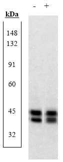
Western Blot for ab17942.
Extracts prepared from PC12 cells not stimulated (-), or stimulated with NGF (+) were resolved by SDS-PAGE on a 10% polyacrylamide gel and transferred to nitrocellulose. Membranes were blocked with a 5% BSA-TBST buffer overnight at 4C, then were incubated with ERK1&2 pan antibody for two hours at room temperature in a 3% BSA-TBST buffer. After washing, membranes were incubated
with goat anti-rabbit IgG alkaline phosphatase.These data show that ab17942 ERK1&2 antibody allows the total amount of ERK1&2 to be measured.
Extracts prepared from PC12 cells not stimulated (-), or stimulated with NGF (+) were resolved by SDS-PAGE on a 10% polyacrylamide gel and transferred to nitrocellulose. Membranes were blocked with a 5% BSA-TBST buffer overnight at 4C, then were incubated with ERK1&2 pan antibody for two hours at room temperature in a 3% BSA-TBST buffer. After washing, membranes were incubated with goat anti-ra


