Anti-CRM1 antibody (ab24189)
Key features and details
- Rabbit polyclonal to CRM1
- Suitable for: IHC-P, ICC/IF, WB
- Reacts with: Human
- Isotype: IgG
Overview
-
Product name
Anti-CRM1 antibody
See all CRM1 primary antibodies -
Description
Rabbit polyclonal to CRM1 -
Host species
Rabbit -
Tested applications
Suitable for: IHC-P, ICC/IF, WBmore details -
Species reactivity
Reacts with: Human -
Immunogen
Synthetic peptide corresponding to Human CRM1 aa 1000 to the C-terminus conjugated to keyhole limpet haemocyanin.
(Peptide available asab25749) -
Positive control
- ab24189 gave a positive result in HeLa whole cell lysate. This antibody also gave a positive signal in IHC in human hippocampus tissue sections.
-
General notes
The Life Science industry has been in the grips of a reproducibility crisis for a number of years. Abcam is leading the way in addressing this with our range of recombinant monoclonal antibodies and knockout edited cell lines for gold-standard validation. Please check that this product meets your needs before purchasing.
If you have any questions, special requirements or concerns, please send us an inquiry and/or contact our Support team ahead of purchase. Recommended alternatives for this product can be found below, along with publications, customer reviews and Q&As
Properties
-
Form
Liquid -
Storage instructions
Shipped at 4°C. Store at +4°C short term (1-2 weeks). Upon delivery aliquot. Store at -20°C or -80°C. Avoid freeze / thaw cycle. -
Storage buffer
pH: 7.40
Preservative: 0.02% Sodium azide
Constituent: PBS
Batches of this product that have a concentration Concentration information loading...
Purity
Immunogen affinity purifiedClonality
PolyclonalIsotype
IgGResearch areas
Associated products
-
Compatible Secondaries
-
Isotype control
-
Recombinant Protein
Applications
The Abpromise guarantee
Our Abpromise guarantee covers the use of ab24189 in the following tested applications.
The application notes include recommended starting dilutions; optimal dilutions/concentrations should be determined by the end user.
Application Abreviews Notes IHC-P Use a concentration of 5 µg/ml. Perform heat mediated antigen retrieval before commencing with IHC staining protocol.ICC/IF (1) Use a concentration of 1 µg/ml.WB (1) Use a concentration of 1 µg/ml. Detects a band of approximately 100 kDa (predicted molecular weight: 123 kDa).Abcam recommends using 3% milk as the blocking agent.
Notes IHC-P
Use a concentration of 5 µg/ml. Perform heat mediated antigen retrieval before commencing with IHC staining protocol.ICC/IF
Use a concentration of 1 µg/ml.WB
Use a concentration of 1 µg/ml. Detects a band of approximately 100 kDa (predicted molecular weight: 123 kDa).Abcam recommends using 3% milk as the blocking agent.
Target
-
Function
Mediates the nuclear export of cellular proteins (cargos) bearing a leucine-rich nuclear export signal (NES) and of RNAs. In the nucleus, in association with RANBP3, binds cooperatively to the NES on its target protein and to the GTPase RAN in its active GTP-bound form (Ran-GTP). Docking of this complex to the nuclear pore complex (NPC) is mediated through binding to nucleoporins. Upon transit of an nuclear export complex into the cytoplasm, disassembling of the complex and hydrolysis of Ran-GTP to Ran-GDP (induced by RANBP1 and RANGAP1, respectively) cause release of the cargo from the export receptor. The directionality of nuclear export is thought to be conferred by an asymmetric distribution of the GTP- and GDP-bound forms of Ran between the cytoplasm and nucleus. Involved in U3 snoRNA transport from Cajal bodies to nucleoli. Binds to late precursor U3 snoRNA bearing a TMG cap. Several viruses, among them HIV-1, HTLV-1 and influenza A use it to export their unspliced or incompletely spliced RNAs out of the nucleus. Interacts with, and mediates the nuclear export of HIV-1 Rev and HTLV-1 Rex proteins. Involved in HTLV-1 Rex multimerization. -
Tissue specificity
Expressed in heart, brain, placenta, lung, liver, skeletal muscle, pancreas, spleen, thymus, prostate, testis, ovary, small intestine, colon and peripheral blood leukocytes. Not expressed in the kidney. -
Sequence similarities
Belongs to the exportin family.
Contains 10 HEAT repeats.
Contains 1 importin N-terminal domain. -
Post-translational
modificationsPhosphorylated upon DNA damage, probably by ATM or ATR. -
Cellular localization
Cytoplasm. Nucleus > nucleoplasm. Nucleus > Cajal body. Nucleus > nucleolus. Located in the nucleoplasm, Cajal bodies and nucleoli. Shuttles between the nucleus/nucleolus and the cytoplasm. - Information by UniProt
-
Database links
- Entrez Gene: 7514 Human
- Omim: 602559 Human
- SwissProt: O14980 Human
- Unigene: 370770 Human
-
Alternative names
- Chromosome region maintenance 1 protein homolog antibody
- CRM 1 antibody
- CRM1 homolog antibody
see all
Images
-
Anti-CRM1 antibody (ab24189) at 1 µg/ml + HeLa (Human epithelial carcinoma cell line) Whole Cell Lysate (ab27252) at 10 µg
Secondary
Goat Anti-Rabbit IgG H&L (HRP) (ab97051) at 1/10000 dilution
Developed using the ECL technique.
Performed under reducing conditions.
Predicted band size: 123 kDa
Observed band size: 120 kDa why is the actual band size different from the predicted?
Additional bands at: 73 kDa (possible non-specific binding)
Exposure time: 90 secondsThis blot was produced using a 4-12% Bis-tris gel under the MOPS buffer system. The gel was run at 200V for 50 minutes before being transferred onto a Nitrocellulose membrane at 30V for 70 minutes. The membrane was then blocked for an hour using 3% milk before being incubated with ab24189 overnight at 4°C. Antibody binding was detected using an anti-rabbit antibody conjugated to HRP, and visualised using ECL development solution.
-
ICC/IF image of ab24189 stained HeLa cells. The cells were 100% methanol fixed (5 min) and then incubated in 1%BSA / 10% normal goat serum / 0.3M glycine in 0.1% PBS-Tween for 1h to permeabilise the cells and block non-specific protein-protein interactions. The cells were then incubated with the antibody ab24189 at 1µg/ml overnight at +4°C. The secondary antibody (green) was DyLight® 488 goat anti- rabbit (ab96899) IgG (H+L) used at a 1/250 dilution for 1h. Alexa Fluor® 594 WGA was used to label plasma membranes (red) at a 1/200 dilution for 1h. DAPI was used to stain the cell nuclei (blue) at a concentration of 1.43µM.
-
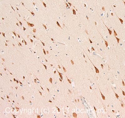
IHC image of CRM1 staining in Human normal hippocampus formalin fixed paraffin embedded tissue section, performed on a Leica Bond™ system using the standard protocol F. The section was pre-treated using heat mediated antigen retrieval with sodium citrate buffer (pH6, epitope retrieval solution 1) for 20 mins. The section was then incubated with ab24189, 5µg/ml, for 15 mins at room temperature and detected using an HRP conjugated compact polymer system. DAB was used as the chromogen. The section was then counterstained with haematoxylin and mounted with DPX.
For other IHC staining systems (automated and non-automated) customers should optimize variable parameters such as antigen retrieval conditions, primary antibody concentration and antibody incubation times.
-
All lanes : Anti-CRM1 antibody (ab24189) at 1 µg/ml
Lane 1 : HeLa whole cell lysate
Lane 2 : HeLa nuclear lysate
Lane 3 : HeLa whole cell lysate with Human CRM1 peptide (ab25749) at 1 µg/ml
Lane 4 : HeLa nuclear cell lysate with Human CRM1 peptide (ab25749) at 1 µg/ml
Lysates/proteins at 20 µg per lane.
Secondary
All lanes : Goat polyclonal to Rabbit IgG at 1/10000 dilution
Performed under reducing conditions.
Predicted band size: 123 kDa
Observed band size: 100 kDa why is the actual band size different from the predicted?ab24189 detects a band of ~ 100 kDa in HeLa whole and HeLa nuclear cell lysates. This is somewhat smaller than the predicted band size according to Swissprot (123 kDa), however as both these bands are specifically blocked by the addition of the immunizing peptide (ab25749) we believe they represent CRM1.
Protocols
Datasheets and documents
-
SDS download
-
Datasheet download
References (22)
ab24189 has been referenced in 22 publications.
- Zhang J et al. The abnormal expression of chromosomal region maintenance 1 (CRM1)-survivin axis in ovarian cancer and its related mechanisms regulating proliferation and apoptosis of ovarian cancer cells. Bioengineered 13:624-633 (2022). PubMed: 34898375
- Lin M et al. Down-regulated expression of CDK5RAP3 and UFM1 suggests a poor prognosis in gastric cancer patients. Front Oncol 12:927751 (2022). PubMed: 36387125
- Zuco V et al. Selinexor versus doxorubicin in dedifferentiated liposarcoma PDXs: evidence of greater activity and apoptotic response dependent on p53 nuclear accumulation and survivin down-regulation. J Exp Clin Cancer Res 40:83 (2021). PubMed: 33648535
- Martini S et al. miR-34a-Mediated Survivin Inhibition Improves the Antitumor Activity of Selinexor in Triple-Negative Breast Cancer. Pharmaceuticals (Basel) 14:N/A (2021). PubMed: 34072442
- Kalita J et al. On the asymmetric partitioning of nucleocytoplasmic transport - recent insights and open questions. J Cell Sci 134:N/A (2021). PubMed: 33912945
Images
-
Anti-CRM1 antibody (ab24189) at 1 µg/ml + HeLa (Human epithelial carcinoma cell line) Whole Cell Lysate (ab27252) at 10 µg
Secondary
Goat Anti-Rabbit IgG H&L (HRP) (ab97051) at 1/10000 dilution
Developed using the ECL technique.
Performed under reducing conditions.
Predicted band size: 123 kDa
Observed band size: 120 kDa why is the actual band size different from the predicted?
Additional bands at: 73 kDa (possible non-specific binding)
Exposure time: 90 secondsThis blot was produced using a 4-12% Bis-tris gel under the MOPS buffer system. The gel was run at 200V for 50 minutes before being transferred onto a Nitrocellulose membrane at 30V for 70 minutes. The membrane was then blocked for an hour using 3% milk before being incubated with ab24189 overnight at 4°C. Antibody binding was detected using an anti-rabbit antibody conjugated to HRP, and visualised using ECL development solution.
-
ICC/IF image of ab24189 stained HeLa cells. The cells were 100% methanol fixed (5 min) and then incubated in 1%BSA / 10% normal goat serum / 0.3M glycine in 0.1% PBS-Tween for 1h to permeabilise the cells and block non-specific protein-protein interactions. The cells were then incubated with the antibody ab24189 at 1µg/ml overnight at +4°C. The secondary antibody (green) was DyLight® 488 goat anti- rabbit (ab96899) IgG (H+L) used at a 1/250 dilution for 1h. Alexa Fluor® 594 WGA was used to label plasma membranes (red) at a 1/200 dilution for 1h. DAPI was used to stain the cell nuclei (blue) at a concentration of 1.43µM.
-
IHC image of CRM1 staining in Human normal hippocampus formalin fixed paraffin embedded tissue section, performed on a Leica Bond™ system using the standard protocol F. The section was pre-treated using heat mediated antigen retrieval with sodium citrate buffer (pH6, epitope retrieval solution 1) for 20 mins. The section was then incubated with ab24189, 5µg/ml, for 15 mins at room temperature and detected using an HRP conjugated compact polymer system. DAB was used as the chromogen. The section was then counterstained with haematoxylin and mounted with DPX.
For other IHC staining systems (automated and non-automated) customers should optimize variable parameters such as antigen retrieval conditions, primary antibody concentration and antibody incubation times.
-
All lanes : Anti-CRM1 antibody (ab24189) at 1 µg/ml
Lane 1 : HeLa whole cell lysate
Lane 2 : HeLa nuclear lysate
Lane 3 : HeLa whole cell lysate with Human CRM1 peptide (ab25749) at 1 µg/ml
Lane 4 : HeLa nuclear cell lysate with Human CRM1 peptide (ab25749) at 1 µg/ml
Lysates/proteins at 20 µg per lane.
Secondary
All lanes : Goat polyclonal to Rabbit IgG at 1/10000 dilution
Performed under reducing conditions.
Predicted band size: 123 kDa
Observed band size: 100 kDa why is the actual band size different from the predicted?ab24189 detects a band of ~ 100 kDa in HeLa whole and HeLa nuclear cell lysates. This is somewhat smaller than the predicted band size according to Swissprot (123 kDa), however as both these bands are specifically blocked by the addition of the immunizing peptide (ab25749) we believe they represent CRM1.


