Anti-C3 antibody (ab129945)
Key features and details
- Rabbit polyclonal to C3
- Suitable for: IHC-P, WB
- Reacts with: Human
- Isotype: IgG
Overview
-
Product name
Anti-C3 antibody
See all C3 primary antibodies -
Description
Rabbit polyclonal to C3 -
Host species
Rabbit -
Tested Applications & Species
See all applications and species dataApplication Species IHC-P HumanWB Human -
Immunogen
Synthetic peptide corresponding to Human C3 aa 1600 to the C-terminus conjugated to keyhole limpet haemocyanin.
Database link: P01024 -
Positive control
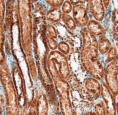
- This antibody gave a positive signal in HepG2 whole cell lysate. This antibody gave a positive result in IHC in the following FFPE tissue: Human normal kidney
Properties
-
Form
Liquid -
Storage instructions
Shipped at 4°C. Store at +4°C short term (1-2 weeks). Upon delivery aliquot. Store at -20°C or -80°C. Avoid freeze / thaw cycle. -
Storage buffer
pH: 7.40
Preservative: 0.02% Sodium azide
Constituent: PBS
Batches of this product that have a concentration Concentration information loading...
Purity
Immunogen affinity purifiedClonality
PolyclonalIsotype
IgGResearch areas
Associated products
-
Compatible Secondaries
-
Isotype control
-
Recombinant Protein
Applications
The Abpromise guarantee
Our Abpromise guarantee covers the use of ab129945 in the following tested applications.
The application notes include recommended starting dilutions; optimal dilutions/concentrations should be determined by the end user.
GuaranteedTested applications are guaranteed to work and covered by our Abpromise guarantee.
PredictedPredicted to work for this combination of applications and species but not guaranteed.
IncompatibleDoes not work for this combination of applications and species.
Application Species IHC-P HumanWB HumanAll applications ChimpanzeeMacaque monkeyGorillaOrangutanApplication Abreviews Notes IHC-P Use a concentration of 5 µg/ml. Perform heat mediated antigen retrieval with citrate buffer pH 6 before commencing with IHC staining protocol.WB Use a concentration of 1 µg/ml. Detects a band of approximately 200 kDa (predicted molecular weight: 187 kDa).Notes IHC-P
Use a concentration of 5 µg/ml. Perform heat mediated antigen retrieval with citrate buffer pH 6 before commencing with IHC staining protocol.WB
Use a concentration of 1 µg/ml. Detects a band of approximately 200 kDa (predicted molecular weight: 187 kDa).Target
-
Function
C3 plays a central role in the activation of the complement system. Its processing by C3 convertase is the central reaction in both classical and alternative complement pathways. After activation C3b can bind covalently, via its reactive thioester, to cell surface carbohydrates or immune aggregates.
Derived from proteolytic degradation of complement C3, C3a anaphylatoxin is a mediator of local inflammatory process. It induces the contraction of smooth muscle, increases vascular permeability and causes histamine release from mast cells and basophilic leukocytes. -
Tissue specificity
Plasma. -
Involvement in disease
Defects in C3 are the cause of complement component 3 deficiency (C3D) [MIM:120700]. A rare defect of the complement classical pathway. Patients develop recurrent, severe, pyogenic infections because of ineffective opsonization of pathogens. Some patients may also develop autoimmune disorders, such as arthralgia and vasculitic rashes, lupus-like syndrome and membranoproliferative glomerulonephritis.
Genetic variation in C3 is associated with susceptibility to age-related macular degeneration type 9 (ARMD9) [MIM:611378]. ARMD is a multifactorial eye disease and the most common cause of irreversible vision loss in the developed world. In most patients, the disease is manifest as ophthalmoscopically visible yellowish accumulations of protein and lipid that lie beneath the retinal pigment epithelium and within an elastin-containing structure known as Bruch membrane.
Defects in C3 are a cause of susceptibility to hemolytic uremic syndrome atypical type 5 (AHUS5) [MIM:612925]. An atypical form of hemolytic uremic syndrome. It is a complex genetic disease characterized by microangiopathic hemolytic anemia, thrombocytopenia, renal failure and absence of episodes of enterocolitis and diarrhea. In contrast to typical hemolytic uremic syndrome, atypical forms have a poorer prognosis, with higher death rates and frequent progression to end-stage renal disease. Note=Susceptibility to the development of atypical hemolytic uremic syndrome can be conferred by mutations in various components of or regulatory factors in the complement cascade system. Other genes may play a role in modifying the phenotype. -
Sequence similarities
Contains 1 anaphylatoxin-like domain.
Contains 1 NTR domain. -
Post-translational
modificationsC3b is rapidly split in two positions by factor I and a cofactor to form iC3b (inactivated C3b) and C3f which is released. Then iC3b is slowly cleaved (possibly by factor I) to form C3c (beta chain + alpha' chain fragment 1 + alpha' chain fragment 2), C3dg and C3f. Other proteases produce other fragments such as C3d or C3g.
Phosphorylation sites are present in the extracelllular medium. -
Cellular localization
Secreted. - Information by UniProt
-
Database links
- Entrez Gene: 718 Human
- Omim: 120700 Human
- SwissProt: P01024 Human
- Unigene: 529053 Human
-
Alternative names
- Acylation stimulating protein cleavage product antibody
- AHUS5 antibody
- ARMD9 antibody
see all
Images
-
IHC image of C3 staining in Human normal Kidney formalin fixed paraffin embedded tissue section, performed on a Leica Bond™ system using the standard protocol F. The section was pre-treated using heat mediated antigen retrieval with sodium citrate buffer (pH6, epitope retrieval solution 1) for 20 mins. The section was then incubated with ab129945, 5µg/ml, for 15 mins at room temperature and detected using an HRP conjugated compact polymer system. DAB was used as the chromogen. The section was then counterstained with haematoxylin and mounted with DPX.
For other IHC staining systems (automated and non-automated) customers should optimize variable parameters such as antigen retrieval conditions, primary antibody concentration and antibody incubation times.
-
Lanes 1 & 3 : Anti-C3 antibody (ab129945) at 1 µg/ml
Lane 2 : Anti-C3 antibody (ab129945) at 1 µg/ml (Milk blocking - 3%)
Lanes 1-2 : HepG2 (Human hepatocellular liver carcinoma cell line) Whole Cell Lysate
Lane 3 : HepG2 (Human hepatocellular liver carcinoma cell line) Whole Cell Lysate with Immunising peptide at 1 µg/ml
Lysates/proteins at 20 µg per lane.
Secondary
All lanes : Goat Anti-Rabbit IgG H&L (HRP) (ab97051) at 1/10000 dilution
Developed using the ECL technique.
Performed under reducing conditions.
Predicted band size: 187 kDa
Observed band size: 200 kDa why is the actual band size different from the predicted?
Additional bands at: 50 kDa (possible non-specific binding), 55 kDa (possible non-specific binding)
Exposure time: 8 minutesThis blot was produced using a 3-8% Tris Acetate gel under the TA buffer system. The gel was run at 150V for 60 minutes before being transferred onto a Nitrocellulose membrane at 30V for 70 minutes. The membrane was then blocked for an hour using 5% Bovine Serum Albumin before being incubated with ab129945 overnight at 4°C. Antibody binding was detected using an anti-rabbit antibody conjugated to HRP, and visualised using ECL development solution.
Protocols
Datasheets and documents
References (2)
ab129945 has been referenced in 2 publications.
- Boire A et al. Complement Component 3 Adapts the Cerebrospinal Fluid for Leptomeningeal Metastasis. Cell 168:1101-1113.e13 (2017). PubMed: 28283064
- Walters MS et al. Smoking accelerates aging of the small airway epithelium. Respir Res 15:94 (2014). IHC-P ; Human . PubMed: 25248511
Images
-
IHC image of C3 staining in Human normal Kidney formalin fixed paraffin embedded tissue section, performed on a Leica Bond™ system using the standard protocol F. The section was pre-treated using heat mediated antigen retrieval with sodium citrate buffer (pH6, epitope retrieval solution 1) for 20 mins. The section was then incubated with ab129945, 5µg/ml, for 15 mins at room temperature and detected using an HRP conjugated compact polymer system. DAB was used as the chromogen. The section was then counterstained with haematoxylin and mounted with DPX.
For other IHC staining systems (automated and non-automated) customers should optimize variable parameters such as antigen retrieval conditions, primary antibody concentration and antibody incubation times.
-
Lanes 1 & 3 : Anti-C3 antibody (ab129945) at 1 µg/ml
Lane 2 : Anti-C3 antibody (ab129945) at 1 µg/ml (Milk blocking - 3%)
Lanes 1-2 : HepG2 (Human hepatocellular liver carcinoma cell line) Whole Cell Lysate
Lane 3 : HepG2 (Human hepatocellular liver carcinoma cell line) Whole Cell Lysate with Immunising peptide at 1 µg/ml
Lysates/proteins at 20 µg per lane.
Secondary
All lanes : Goat Anti-Rabbit IgG H&L (HRP) (ab97051) at 1/10000 dilution
Developed using the ECL technique.
Performed under reducing conditions.
Predicted band size: 187 kDa
Observed band size: 200 kDa why is the actual band size different from the predicted?
Additional bands at: 50 kDa (possible non-specific binding), 55 kDa (possible non-specific binding)
Exposure time: 8 minutesThis blot was produced using a 3-8% Tris Acetate gel under the TA buffer system. The gel was run at 150V for 60 minutes before being transferred onto a Nitrocellulose membrane at 30V for 70 minutes. The membrane was then blocked for an hour using 5% Bovine Serum Albumin before being incubated with ab129945 overnight at 4°C. Antibody binding was detected using an anti-rabbit antibody conjugated to HRP, and visualised using ECL development solution.


