Anti-c-Fos (phospho T325) antibody (ab27793)
Key features and details
- Rabbit polyclonal to c-Fos (phospho T325)
- Suitable for: ICC/IF, ChIP, WB
- Reacts with: Human
- Isotype: IgG
Overview
-
Product name
Anti-c-Fos (phospho T325) antibody
See all c-Fos primary antibodies -
Description
Rabbit polyclonal to c-Fos (phospho T325) -
Host species
Rabbit -
Specificity
ab27793 recognises the cFos phosphorilated at threonine 325 form. -
Tested applications
Suitable for: ICC/IF, ChIP, WBmore details -
Species reactivity
Reacts with: Human -
Immunogen
Synthetic phosphopeptide derived from the region of human cFos that contains threonine 325.
Properties
-
Form
Liquid -
Storage instructions
Shipped at 4°C. Store at +4°C short term (1-2 weeks). Upon delivery aliquot. Store at -20°C long term. -
Storage buffer
pH: 7.30
Preservative: 0.05% Sodium azide
Constituents: PBS, 50% Glycerol (glycerin, glycerine), 0.1% BSA -
 Concentration information loading...
 -
Purity
Immunogen affinity purified -
Purification notes
ab27793 was negatively preadsorbed using a non phosphopeptide corresponding to the site of phosphorylation to remove antibody that is reactive with non phosphorylated cFos. Immunogen affinity purification followed. -
Clonality
Polyclonal -
Isotype
IgG -
Research areas
Images
-
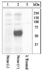
All lanes : Anti-c-Fos (phospho T325) antibody (ab27793) at 1/1000 dilution (diluted in a 3% Milk TBST buffer.)
Lane 1 : Non EGF treated A431 cell lysate
Lane 2 : EGF treated A431 cell lysate
Lane 3 : EGF treated A431 cell lysate treated with Lambda phosphatase
Secondary
All lanes : goat F(ab’)2 anti rabbit IgG HRP conjugate
Predicted band size: 41 kDa
Observed band size: 58 kDa why is the actual band size different from the predicted?
The figure shows that the phosphorylation of cFos on threonine 325 is induced by EGF treatment and that Lambda phosphatase treatment eliminates the signal, thereby demonstrating the phospho specificity of ab27793. -
Immunofluorescence analysis of c-Fos (phospho T325) was done on 70% confluent log phase HeLa cell treated with 200 nM of PMA for 20 minutes. The cell were fixed with 4% paraformaldehyde for 10 minutes, permeabilized with 0.1% Triton™ X-100 for 10 minutes, and blocked with 1% BSA for 1 hour at room temperature. The cells were labeled with Phosc-Fos (phospho T325)Rabbit Polyclonal Antibody (ab27793) at 2 ug/ml in 0.1% BSA and incubated for 3 hours at room temperature and then labeled with Goat anti-Rabbit IgG (H+L) Secondary Antibody, Alexa Fluor® 488 conjugate at a dilution of 1/2000 for 45 minutes at room temperature (Panel a: green). Nuclei (Panel b: blue) were stained with DAPI. F-actin (Panel c: red) was stained with Alexa Fluor® 555 Rhodamine Phalloidin. Panel d is a merged image showing nuclear and cytoplasmic localization. Panel e is untreated cell with no signal. Panel f represents control cells with no primary antibody to assess background. The images were captured at 60X magnification.
-
Chromatin Immunoprecipitation (ChIP) was performed using Anti-c-Fos (phospho T325) antibody (ab27793) 3 ug on sheared chromatin from 2 million A431 cells treated with EGF (200ng/ml), for 30 minutes. Normal Rabbit IgG was used as a negative IP control. The purified DNA was analyzed by 7500 Fast qPCR system with optimized PCR primer pairs for the promoter of active IL-6, CDKN1A gene, used as positive control target, and the SAT2, used as negative control target. Data is presented as fold enrichment of the antibody signal versus the negative control IgG using the comparative CT method.


