Anti-NeuN antibody - Neuronal Marker (ab104225)
Key features and details
- Rabbit polyclonal to NeuN - Neuronal Marker
- Suitable for: WB, IHC-FrFl, IHC-P, IHC-FoFr
- Reacts with: Mouse, Rat, Human
- Isotype: IgG
Overview
-
Product name
Anti-NeuN antibody - Neuronal Marker
See all NeuN primary antibodies -
Description
Rabbit polyclonal to NeuN - Neuronal Marker -
Host species
Rabbit -
Tested Applications & Species
See all applications and species dataApplication Species IHC-FoFr RatIHC-FrFl MouseIHC-P MouseRatHumanWB Mouse -
Immunogen
Synthetic peptide. This information is proprietary to Abcam and/or its suppliers.
-
General notes
We are constantly working hard to ensure we provide our customers with best in class antibodies. As a result of this work we are pleased to now offer this antibody in purified format. We are in the process of updating our datasheets. The purified format is designated 'PUR' on our product labels. If you have any questions regarding this update, please contact our Scientific Support team.
Properties
-
Form
Liquid -
Storage instructions
Shipped at 4°C. Store at +4°C short term (1-2 weeks). Upon delivery aliquot. Store at -20°C. Avoid freeze / thaw cycle. -
Storage buffer
Preservative: 0.03% Sodium azide
Constituents: 49.98% PBS, 49.98% Glycerol -
 Concentration information loading...
 -
Purity
Affinity purified -
Clonality
Polyclonal -
Isotype
IgG -
Research areas
Images
-
 Immunohistochemistry (Formalin/PFA-fixed paraffin-embedded sections) - Anti-NeuN antibody - Neuronal Marker (ab104225)
Immunohistochemical analysis of paraformaldehyde fixed frozen section Rat hippocampus tissue labeling NeuN with ab104225 at 1/1000 dilution.
-
 Immunohistochemistry (Formalin/PFA-fixed paraffin-embedded sections) - Anti-NeuN antibody - Neuronal Marker (ab104225)
IHC image of NeuN staining in rat cerebellum formalin fixed paraffin embedded tissue section, performed on a Leica Bond™ system using the standard protocol F. The section was pre-treated using heat mediated antigen retrieval with sodium citrate buffer (pH6, epitope retrieval solution 1) for 20 mins. The section was then incubated with ab104225, 1in500 dilution, for 15 mins at room temperature and detected using an HRP conjugated compact polymer system. DAB was used as the chromogen. The section was then counterstained with haematoxylin and mounted with DPX.
For other IHC staining systems (automated and non-automated) customers should optimize variable parameters such as antigen retrieval conditions, primary antibody concentration and antibody incubation times.
-
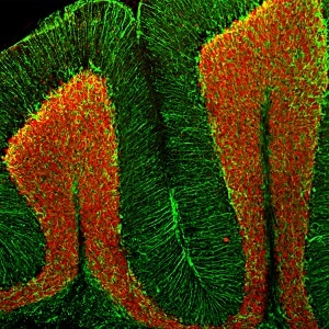
Immunofluorescent analysis of a section of adult mouse cerebellum stained with ab104225 at a dilution 1:5,000 in red, co-stained with chicken pAb to GFAP, dilution 1:5,000, in green.
Following transcardial perfusion of mouse with 4% paraformaldehyde, brain was post fixed for 24 hours, cut to 45 μM, and free-floating sections. ab104225 stains the nuclei of neurons in the cerebellar granule layer. The GFAP antibody stains the processes of Bergmann glia in the molecular layer and astroglia in the granule and white matter layers.
-
All lanes : Anti-NeuN antibody - Neuronal Marker (ab104225) at 1/1000 dilution
Lane 2 : Mouse whole brain lysate
Lane 3 : Rat whole brain lysate
Predicted band size: 48 kDa
-
 Immunohistochemistry (Formalin/PFA-fixed paraffin-embedded sections) - Anti-NeuN antibody - Neuronal Marker (ab104225)
IHC image of NeuN staining in human cerebellum formalin fixed paraffin embedded tissue section, performed on a Leica Bond™ system using the standard protocol F. The section was pre-treated using heat mediated antigen retrieval with sodium citrate buffer (pH6, epitope retrieval solution 1) for 20 mins. The section was then incubated with ab104225, 1in500 dilution, for 15 mins at room temperature and detected using an HRP conjugated compact polymer system. DAB was used as the chromogen. The section was then counterstained with haematoxylin and mounted with DPX.
For other IHC staining systems (automated and non-automated) customers should optimize variable parameters such as antigen retrieval conditions, primary antibody concentration and antibody incubation times.
-
 Immunohistochemistry (Formalin/PFA-fixed paraffin-embedded sections) - Anti-NeuN antibody - Neuronal Marker (ab104225)
IHC image of NeuN staining in mouse cerebellum formalin fixed paraffin embedded tissue section, performed on a Leica Bond™ system using the standard protocol B. The section was pre-treated using heat mediated antigen retrieval with sodium citrate buffer (pH6, epitope retrieval solution 1) for 20 mins. The section was then incubated with ab104225, 1in500 dilution, for 15 mins at room temperature. A goat anti-rabbit biotinylated secondary antibody was used to detect the primary, and visualized using an HRP conjugated ABC system. DAB was used as the chromogen. The section was then counterstained with haematoxylin and mounted with DPX.
For other IHC staining systems (automated and non-automated) customers should optimize variable parameters such as antigen retrieval conditions, primary antibody concentration and antibody incubation times.
-
 Immunohistochemistry (Formalin/PFA-fixed paraffin-embedded sections) - Anti-NeuN antibody - Neuronal Marker (ab104225) This image is courtesy of an abreview submitted by Carl Hobbs, King's College London, United Kingdom
IHC-P image of FOX3/NeuN staining using rat DRG using ab104225 (1:500). The sections were subjected to heat mediated antigen retrieval using citric acid pH 6. The sections were blocked using 1% BSA for 10 mins at 21°C. ab104225 was incubated for 2 hours at 21°C. The secondary antibody used was Goat polyclonal to Rabbit IgG conjugated to biotin (1:200).
-
 Immunohistochemistry (PFA perfusion fixed frozen sections) - Anti-NeuN antibody - Neuronal Marker (ab104225)
ab104225 at 1/1000 dilution, staining FOX3 (red) in rat brain cortex and counterstained for DNA in blue.
-
 Immunohistochemistry (Formalin/PFA-fixed paraffin-embedded sections) - Anti-NeuN antibody - Neuronal Marker (ab104225) This image is courtesy of an abreview submitted by Carl Hobbs, King's College London, United Kingdom
IHC-P image of FOX3/NeuN staining using mouse DRG using ab104225 (1:500). The sections were subjected to heat mediated antigen retrieval using citric acid pH 6. The sections were blocked using 1% BSA for 10 mins at 21°C. ab104225 was incubated for 2 hours at 21°C. The secondary antibody used was Goat polyclonal to Rabbit IgG conjugated to biotin (1:200).


