Anti-BACE1 antibody (ab2077)
Key features and details
- Rabbit polyclonal to BACE1
- Suitable for: WB, ICC, IHC-P
- Reacts with: Mouse, Human
- Isotype: IgG
Overview
-
Product name
Anti-BACE1 antibody
See all BACE1 primary antibodies -
Description
Rabbit polyclonal to BACE1 -
Host species
Rabbit -
Tested applications
Suitable for: WB, ICC, IHC-Pmore details -
Species reactivity
Reacts with: Mouse, Human -
Immunogen
-
Positive control
- WB: Human A431, A549, Caco-2, Daudi, HeLa, K562, Jurkat, SK-N-SH, THP-1 and brain tissue lysates. Mouse: 3T3/NIH cell lysate. IHC-P: Mouse brain tissue. ICC: Mouse 3T3 cells.
-
General notes
Beta-site APP Cleaving Enzyme.
The Life Science industry has been in the grips of a reproducibility crisis for a number of years. Abcam is leading the way in addressing this with our range of recombinant monoclonal antibodies and knockout edited cell lines for gold-standard validation. Please check that this product meets your needs before purchasing.
If you have any questions, special requirements or concerns, please send us an inquiry and/or contact our Support team ahead of purchase. Recommended alternatives for this product can be found below, along with publications, customer reviews and Q&As
Properties
-
Form
Liquid -
Storage instructions
Shipped at 4°C. Store at +4°C short term (1-2 weeks). Upon delivery aliquot. Store at -20°C. Avoid freeze / thaw cycle. Stable for 12 months at -20°C. -
Storage buffer
pH: 7.2
Preservative: 0.02% Sodium azide -
 Concentration information loading...
 -
Purity
Ion Exchange Chromatography -
Purification notes
BACE Antibody is Ion exchange chromatography purified. -
Primary antibody notes
Beta-site APP Cleaving Enzyme. -
Clonality
Polyclonal -
Isotype
IgG -
Research areas
Images
-
All lanes :
Lane 1 : A431 (Human epidermoid carcinoma cell line) whole cell lysate withAnti-BACE1 antibody (ab2077)
Lane 2 : A549 (Human lung carcinoma cell line) whole cell lysate withAnti-BACE1 antibody (ab2077)
Lane 3 : Caco-2 (Human colorectal adenocarcinoma cell line) whole cell lysate withAnti-BACE1 antibody (ab2077)
Lane 4 : Daudi (Human Burkitt's lymphoma cell line) whole cell lysate withAnti-BACE1 antibody (ab2077)
Lane 5 : HeLa (Human epithelial cell line from cervix adenocarcinoma) whole cell lysate withAnti-BACE1 antibody (ab2077)
Lane 6 : K562 (Human chronic myelogenous leukemia cell line from bone marrow ) whole cell lysate withAnti-BACE1 antibody (ab2077)
Lane 7 : Jurkat (Human T cell leukemia cell line from peripheral blood) whole cell lysate withAnti-BACE1 antibody (ab2077)
Lane 8 : SK-N-SH (Human neuroblastoma cell line) whole cell lysate withAnti-BACE1 antibody (ab2077)
Lane 9 : THP-1 (Human monocytic leukemia cell line) whole cell lysate withAnti-BACE1 antibody (ab2077)
Lysates/proteins at 15 µg per lane.
Blocking peptides at 1 µg/ml per lane.
Secondary
All lanes : Rabbit IgG antibody (HRP) at 1/10000 dilution
Developed using the ECL technique.
Additional bands at: 65 kDa (possible glycosylated form)10% gel.
Running conditions: 130v for 2 hours.
Transfer conditions: wet, 250mA, 2 hrs (Nitrocellulose membrane).
Blocking condition: 5% non-fat dry milk in TBS, 4C, overnight.
Primary antibody incubation: Room temperature for 1 hour.
Secondary antibody incubation: Room temperature for 1 hour.
Washing conditions: 15 mL TSBT, 3 x 10 minutes.
Exposure: ECL solution
-
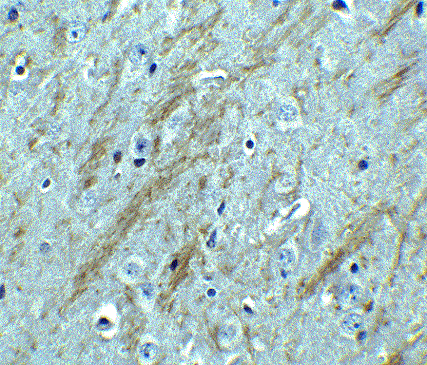
Immunohistochemical analysis of paraffin-embedded mouse brain tissue using ab2077 at 2.5 µg/ml. Tissue was fixed with formaldehyde and blocked with 10% serum for 1 h at RT; antigen retrieval was by heat mediation with a citrate buffer (pH6). Samples were incubated with primary antibody overnight at 4°C. A goat anti-rabbit IgG H&L (HRP) at 1/250 was used as secondary. Counter stained with Hematoxylin.
-
Immunofluorescent analysis of 4% paraformaldehyde-fixed NIH/3T3 (Mouse embryo fibroblast cell line) cells labeling BACE1 with ab2077 at 20 ug/mL, followed by goat anti-rabbit IgG secondary antibody at 1/500 dilution (green) and DAPI staining (blue). Image showing both membrane and cytosol staining on NIH/3T3 cells.
-
Immunocytochemistry/ Immunofluorescence analysis of NIH/3T3 (Mouse embryo fibroblast cell line) cells labeling BACE1 with ab2077 at 10 μg/mL. Cells were fixed with formaldehyde and blocked with 10% serum for 1 h at RT; antigen retrieval was by heat mediation with a citrate buffer (pH6). Samples were incubated with primary antibody overnight at 4oC. A goat anti-rabbit IgG H&L (HRP) at 1/250 was used as secondary. Counter stained with Hematoxylin.
-
All lanes : Anti-BACE1 antibody (ab2077) at 1 µg/ml
Lane 1 : Human brain tissue lysate with absence of blocking peptide
Lane 2 : Human brain tissue
lysate with BACE1 peptide (ab7883)
Lane 3 : Mouse 3T3/NIH cell lysate
Lysates/proteins at 15 µg per lane.
Secondary
All lanes : Goat anti-rabbit IgG HRP conjugate at 1/10000 dilution
Observed band size: 70 kDa why is the actual band size different from the predicted?Incubate the antibody for 1 hour at room temperature in 5% NFDM/TBST.
-
 Immunocytochemistry/ Immunofluorescence - Anti-BACE1 antibody (ab2077) Image courtesy of an anonymous Abreview.
Immunocytochemiscal analysis of D54MG (human glioblastoma cell line) cells labeling BACE1 with ab2077.Cells were fixed in paraformaldehyde, permeabilized with 0.1% Triton X-100, blocked with 0.5% BSA for 20 minutes at room temperature, then incubated with ab2077 at a 1/50 dilution for 16 hours at 4°C. The secondary used was a TRITC conjugated goat anti-rabbit polyclonal, used at a 1/400 dilution. Nuclei are counterstained with DAPI.


