Human MIP1a ELISA Kit, Fluorescent (ab229400)
")
Key features and details
- One-wash 90 minute protocol
- Sensitivity: 0.2 pg/ml
- Range: 0.2 pg/ml - 200 pg/ml
- Sample type: Cell culture supernatant, Cit plasma, EDTA Plasma, Hep Plasma, Serum
- Detection method: Fluorescent
- Assay type: Sandwich (quantitative)
- Reacts with: Human
Overview
-
Product name
Human MIP1a ELISA Kit, Fluorescent
See all Macrophage Inflammatory Protein 1 alpha / CCL3 kits -
Detection method
Fluorescent -
Precision
Intra-assay Sample n Mean SD CV% Supernatant 5 1.9% Inter-assay Sample n Mean SD CV% Supernatant 3 7.2% -
Sample type
Cell culture supernatant, Serum, Hep Plasma, EDTA Plasma, Cit plasma -
Assay type
Sandwich (quantitative) -
Sensitivity
0.2 pg/ml -
Range
0.2 pg/ml - 200 pg/ml -
Recovery
Sample specific recovery Sample type Average % Range Cell culture supernatant 101 100% - 102% Serum 99 96% - 101% Hep Plasma 97 94% - 100% EDTA Plasma 100 92% - 105% Cit plasma 95 91% - 101% -
Assay time
1h 30m -
Assay duration
One step assay -
Species reactivity
Reacts with: Human
Does not react with: Cow -
Product overview
MIP1a in vitro CatchPoint® SimpleStep ELISA® (Enzyme-Linked Immunosorbent Assay) kit is designed for the quantitative measurement of MIP1a protein in human serum, plasma and cell culture supernatant.
This CatchPoint SimpleStep ELISA kit has been optimized for Molecular Devices Microplate Readers. Click here for a list of recommended Microplate Readers.
If using a Molecular Devices’ plate reader supported by SoftMax® Pro software, a preconfigured protocol for these CatchPoint SimpleStep ELISA Kits is available with all the protocol and analysis settings at www.softmaxpro.org.The CatchPoint® SimpleStep ELISA® employs an affinity tag labeled capture antibody and a reporter conjugated detector antibody which immunocapture the sample analyte in solution. This entire complex (capture antibody/analyte/detector antibody) is in turn immobilized via immunoaffinity of an anti-tag antibody coating the well. To perform the assay, samples or standards are added to the wells, followed by the antibody mix. After incubation, the wells are washed to remove unbound material. CatchPoint® HRP Development Solution containing the Stoplight Red Substrate is added. During incubation, the substrate is catalyzed by HRP generating a fluorescent product. Signal is generated proportionally to the amount of bound analyte and the intensity is measured in a fluorescence plater reader at 530/570/590 nm Excitation/Cutoff/Emission.
16% reactivity observed in mouse.
-
Notes
Macrophage Inflammatory Protein 1-alpha (MIP1a, also known as CCL3) is a monokine with both inflammatory and chemokine properties. MIP-1-alpha can bind to CCR1, CCR4 and CCR5. In addition, it is one of the major HIV-suppressive factors produced by CD8+ T-cells. Recombinant MIP-1-alpha induces a dose-dependent inhibition of different strains of HIV-1, HIV-2, and simian immunodeficiency virus (SIV).
-
Platform
Pre-coated microplate (12 x 8 well strips)
Properties
-
Storage instructions
Store at +4°C. Please refer to protocols. -
Components 1 x 96 tests 100X Stoplight Red Substrate 1 x 120µl 10X Human MIP1a Capture Antibody 1 x 600µl 10X Human MIP1a Detector Antibody 1 x 600µl 10X Wash Buffer PT (ab206977) 1 x 20ml 500X Hydrogen Peroxide (H2O2, 3%) 1 x 50µl Antibody Diluent CPI - HAMA Blocker (ab193969) 1 x 6ml Human MIP1a Lyophilized Recombinant Protein 2 vials Plate Seals 1 unit Sample Diluent NS (ab193972) 1 x 50ml SimpleStep Pre-Coated Black 96-Well Microplate 1 unit Stoplight Red Substrate Buffer 1 x 12ml -
Research areas
-
Function
Monokine with inflammatory and chemokinetic properties. Binds to CCR1, CCR4 and CCR5. One of the major HIV-suppressive factors produced by CD8+ T-cells. Recombinant MIP-1-alpha induces a dose-dependent inhibition of different strains of HIV-1, HIV-2, and simian immunodeficiency virus (SIV). -
Sequence similarities
Belongs to the intercrine beta (chemokine CC) family. -
Post-translational
modificationsN-terminal processed form LD78-alpha(4-69) is produced by proteolytic cleavage after secretion from HTLV1-transformed T-cells. -
Cellular localization
Secreted. - Information by UniProt
-
Alternative names
- C C motif chemokine 3
- CCL 3
- CCL3
see all -
Database links
- Entrez Gene: 6348 Human
- Omim: 182283 Human
- SwissProt: P10147 Human
- Unigene: 514107 Human
Images
-
Other - Human MIP1a ELISA Kit, Fluorescent (ab229400)
SimpleStep ELISA technology allows the formation of the antibody-antigen complex in one single step, reducing assay time to 90 minutes. Add samples or standards and antibody mix to wells all at once, incubate, wash, and add your final substrate. See protocol for a detailed step-by-step guide.
-
 Example of human MIP1a standard curve in Sample Diluent NS.
The MIP1a standard curve was prepared as described in Section 10. Background-subtracted data values (mean +/- SD) are graphed.
-
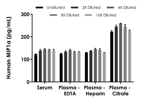 Interpolated concentrations of spiked MIP1a in human serum and plasma samples.
The concentrations of MIP1a were measured in duplicates, interpolated from the MIP1a standard curves and corrected for sample dilution. Undiluted samples are as follows: serum 50%, plasma (EDTA) 50%, plasma (heparin) 50%) and plasma (citrate) 25%. The interpolated dilution factor corrected values are plotted (mean +/- SD, n=2).
-
 Serum from ten individual healthy human female donors was measured in duplicate.
Interpolated dilution factor corrected values are plotted (mean +/- SD, n=2). The mean MIP1a concentration was determined to be 5.5 pg/mL with a range of 1.3 – 12.1 pg/mL.
-
 Interpolated concentrations of native MIP1a in human peripheral blood monocyte (PBMC) cell culture supernatant samples.
The concentrations of MIP1a were measured in duplicates, interpolated from the MIP1a standard curves and corrected for sample dilution. Undiluted samples are as follows: unstimulated 6.35% and stimulated 0.2%. The interpolated dilution factor corrected values are plotted (mean +/- SD, n=2). The mean MIP1a concentration was determined to be 0.241 ng/mL in unstimulated PBMC cell culture supernatant and 40.8 ng/mL in stimulated.
-
 Sandwich ELISA - Human MIP1a ELISA Kit, Fluorescent (ab229400)To learn more about the advantages of recombinant antibodies see here.


