Anti-Transglutaminase 2 antibody (ab64771)
")
Key features and details
- Rabbit polyclonal to Transglutaminase 2
- Suitable for: IP, IHC-P, ICC/IF, WB
- Knockout validated
- Reacts with: Human
- Isotype: IgG
Overview
-
Product name
Anti-Transglutaminase 2 antibody
See all Transglutaminase 2 primary antibodies -
Description
Rabbit polyclonal to Transglutaminase 2 -
Host species
Rabbit -
Tested Applications & Species
See all applications and species dataApplication Species ICC/IF HumanIHC-P HumanIP HumanWB Human
-
Immunogen
Synthetic peptide conjugated to KLH derived from within residues 600 to the C-terminus of Human Transglutaminase 2.
Read Abcam's proprietary immunogen policy (Peptide available as ab73168.) -
Positive control
- This antibody gave a positive signal in the following Human Lysates: Placenta Tissue, HeLa Whole Cell - Staurosporine Treated (24hr, 500nM), HeLa Whole Cell
Properties
-
Form
Liquid -
Storage instructions
Shipped at 4°C. Store at +4°C short term (1-2 weeks). Upon delivery aliquot. Store at -20°C or -80°C. Avoid freeze / thaw cycle. -
Storage buffer
pH: 7.40
Preservative: 0.02% Sodium azide
Constituent: PBS
Batches of this product that have a concentration Concentration information loading...
Purity
Immunogen affinity purifiedClonality
PolyclonalIsotype
IgGResearch areas
Associated products
-
Compatible Secondaries
-
Isotype control
-
Recombinant Protein
Applications
The Abpromise guarantee
Our Abpromise guarantee covers the use of ab64771 in the following tested applications.
The application notes include recommended starting dilutions; optimal dilutions/concentrations should be determined by the end user.
GuaranteedTested applications are guaranteed to work and covered by our Abpromise guarantee.
PredictedPredicted to work for this combination of applications and species but not guaranteed.
IncompatibleDoes not work for this combination of applications and species.
Application Species ICC/IF HumanIHC-P HumanIP HumanWB HumanAll applications CowChimpanzeeApplication Abreviews Notes IP Use a concentration of 5 µg/ml.IHC-P Use a concentration of 1 µg/ml.ICC/IF Use a concentration of 5 µg/ml.WB Use a concentration of 1 µg/ml. Detects a band of approximately 77 kDa (predicted molecular weight: 77 kDa).Notes IP
Use a concentration of 5 µg/ml.IHC-P
Use a concentration of 1 µg/ml.ICC/IF
Use a concentration of 5 µg/ml.WB
Use a concentration of 1 µg/ml. Detects a band of approximately 77 kDa (predicted molecular weight: 77 kDa).Target
-
Function
Catalyzes the cross-linking of proteins and the conjugation of polyamines to proteins. -
Sequence similarities
Belongs to the transglutaminase superfamily. Transglutaminase family. - Information by UniProt
-
Database links
- Entrez Gene: 281528 Cow
- Entrez Gene: 7052 Human
- Omim: 190196 Human
- SwissProt: P51176 Cow
- SwissProt: P21980 Human
- Unigene: 517033 Human
-
Alternative names
- ALPHA SUBUNIT antibody
- C polypeptide antibody
- EC 2.3.2.13 antibody
see all
Images
-
Western blot - Anti-Transglutaminase 2 antibody (ab64771)All lanes : Anti-Transglutaminase 2 antibody (ab64771) at 1/5000 dilution
Lane 1 : HeLa whole cell lysate
Lane 2 : Huvec whole cell lysate
Lane 3 : Wild-type A549 whole cell lysate
Lane 4 : TGM2 knockout A549 whole cell lysate
Lysates/proteins at 20 µg per lane.
Predicted band size: 77 kDa
Observed band size: 77 kDaLanes 1 - 4: Merged signal (red and green). Green - ab64771 observed at 77 kDa. Red - loading control, ab8245, observed at 37 kDa.
ab64771 was shown to specifically react with in wild-type A549 cells as signal was lost in TGM2 knockout cells. Wild-type and TGM2 knockout samples were subjected to SDS-PAGE. Ab64771 and ab8245 (Mouse anti GAPDH loading control) were incubated overnight at 4°C at 1/5000 dilution and 1/20000 dilution respectively. Blots were developed with Goat anti-Rabbit IgG H&L (IRDye® 800CW) preabsorbed ab216773 and Goat anti-Mouse IgG H&L (IRDye® 680RD) preabsorbed ab216776 secondary antibodies at 1/20000 dilution for 1 hour at room temperature before imaging.
-
 Western blot - Anti-Transglutaminase 2 antibody (ab64771)All lanes : Anti-Transglutaminase 2 antibody (ab64771) at 1 µg/ml
Lane 1 : Human placenta tissue lysate - total protein (ab29745)
Lane 2 : HeLa Whole Cell Lysate - Staurosporine Treated (24hr, 500nM)
Lane 3 : HeLa (Human epithelial carcinoma cell line) Whole Cell Lysate
Lysates/proteins at 10 µg per lane.
Secondary
All lanes : Goat polyclonal to Rabbit IgG - H&L - Pre-Adsorbed (HRP) at 1/3000 dilution
Developed using the ECL technique.
Performed under reducing conditions.
Predicted band size: 77 kDa
Observed band size: 77 kDa
Exposure time: 30 seconds -
 Immunoprecipitation - Anti-Transglutaminase 2 antibody (ab64771)
Transglutaminase 2 was immunoprecipitated using 0.5mg Hela whole cell extract, 5µg of Rabbit polyclonal to Transglutaminase 2 and 50µl of protein G magnetic beads (+). No antibody was added to the control (-).
The antibody was incubated under agitation with Protein G beads for 10min, Hela whole cell extract lysate diluted in RIPA buffer was added to each sample and incubated for a further 10min under agitation.
Proteins were eluted by addition of 40µl SDS loading buffer and incubated for 10min at 70°C; 10µl of each sample was separated on a SDS PAGE gel, transferred to a nitrocellulose membrane, blocked with 5% BSA and probed with ab64771.
Secondary: Mouse monoclonal [SB62a] Secondary Antibody to Rabbit IgG light chain (HRP) (ab99697).
Band: 77kDa; Transglutaminase 2, non specific - as present in control (lane 2); 75kDa: We are unsure as to the identity of this extra band.
-
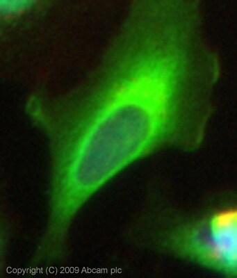 Immunocytochemistry/ Immunofluorescence - Anti-Transglutaminase 2 antibody (ab64771)ICC/IF image of ab64771 stained HeLa cells. The cells were 4% PFA fixed (10 min) and then incubated in 1%BSA / 10% normal goat serum / 0.3M glycine in 0.1% PBS-Tween for 1h to permeabilise the cells and block non-specific protein-protein interactions. The cells were then incubated with the antibody (ab64771, 5µg/ml) overnight at +4°C. The secondary antibody (green) was Alexa Fluor® 488 goat anti-rabbit IgG (H+L) used at a 1/1000 dilution for 1h. Alexa Fluor® 594 WGA was used to label plasma membranes (red) at a 1/200 dilution for 1h. DAPI was used to stain the cell nuclei (blue) at a concentration of 1.43µM. This antibody also gave a positive result in 4% PFA fixed (10 min) Hek293, HepG2 and MCF7 cells at 5µg/ml, and in 100% methanol fixed (5 min) HeLa, Hek293, HepG2 and MCF7 cells at 5µg/ml
 -
 Immunohistochemistry (Formalin/PFA-fixed paraffin-embedded sections) - Anti-Transglutaminase 2 antibody (ab64771)IHC image of Transglutaminase 2 staining in Human Tonsil FFPE section, performed on a BondTM system using the standard protocol F. The section was pre-treated using heat mediated antigen retrieval with sodium citrate buffer (pH6, epitope retrieval solution 1) for 20 mins. The section was then incubated with ab64771, 1µg/ml, for 15 mins at room temperature and detected using an HRP conjugated compact polymer system. DAB was used as the chromogen. The section was then counterstained with haematoxylin and mounted with DPX
Protocols
Datasheets and documents
References (0)
ab64771 has not yet been referenced specifically in any publications.
Images
-
Western blot - Anti-Transglutaminase 2 antibody (ab64771)All lanes : Anti-Transglutaminase 2 antibody (ab64771) at 1/5000 dilution
Lane 1 : HeLa whole cell lysate
Lane 2 : Huvec whole cell lysate
Lane 3 : Wild-type A549 whole cell lysate
Lane 4 : TGM2 knockout A549 whole cell lysate
Lysates/proteins at 20 µg per lane.
Predicted band size: 77 kDa
Observed band size: 77 kDaLanes 1 - 4: Merged signal (red and green). Green - ab64771 observed at 77 kDa. Red - loading control, ab8245, observed at 37 kDa.
ab64771 was shown to specifically react with in wild-type A549 cells as signal was lost in TGM2 knockout cells. Wild-type and TGM2 knockout samples were subjected to SDS-PAGE. Ab64771 and ab8245 (Mouse anti GAPDH loading control) were incubated overnight at 4°C at 1/5000 dilution and 1/20000 dilution respectively. Blots were developed with Goat anti-Rabbit IgG H&L (IRDye® 800CW) preabsorbed ab216773 and Goat anti-Mouse IgG H&L (IRDye® 680RD) preabsorbed ab216776 secondary antibodies at 1/20000 dilution for 1 hour at room temperature before imaging.
-
Western blot - Anti-Transglutaminase 2 antibody (ab64771)All lanes : Anti-Transglutaminase 2 antibody (ab64771) at 1 µg/ml
Lane 1 : Human placenta tissue lysate - total protein (ab29745)
Lane 2 : HeLa Whole Cell Lysate - Staurosporine Treated (24hr, 500nM)
Lane 3 : HeLa (Human epithelial carcinoma cell line) Whole Cell Lysate
Lysates/proteins at 10 µg per lane.
Secondary
All lanes : Goat polyclonal to Rabbit IgG - H&L - Pre-Adsorbed (HRP) at 1/3000 dilution
Developed using the ECL technique.
Performed under reducing conditions.
Predicted band size: 77 kDa
Observed band size: 77 kDa
Exposure time: 30 seconds
-
Immunoprecipitation - Anti-Transglutaminase 2 antibody (ab64771)
Transglutaminase 2 was immunoprecipitated using 0.5mg Hela whole cell extract, 5µg of Rabbit polyclonal to Transglutaminase 2 and 50µl of protein G magnetic beads (+). No antibody was added to the control (-).
The antibody was incubated under agitation with Protein G beads for 10min, Hela whole cell extract lysate diluted in RIPA buffer was added to each sample and incubated for a further 10min under agitation.
Proteins were eluted by addition of 40µl SDS loading buffer and incubated for 10min at 70°C; 10µl of each sample was separated on a SDS PAGE gel, transferred to a nitrocellulose membrane, blocked with 5% BSA and probed with ab64771.
Secondary: Mouse monoclonal [SB62a] Secondary Antibody to Rabbit IgG light chain (HRP) (ab99697).
Band: 77kDa; Transglutaminase 2, non specific - as present in control (lane 2); 75kDa: We are unsure as to the identity of this extra band.
-
Immunocytochemistry/ Immunofluorescence - Anti-Transglutaminase 2 antibody (ab64771)ICC/IF image of ab64771 stained HeLa cells. The cells were 4% PFA fixed (10 min) and then incubated in 1%BSA / 10% normal goat serum / 0.3M glycine in 0.1% PBS-Tween for 1h to permeabilise the cells and block non-specific protein-protein interactions. The cells were then incubated with the antibody (ab64771, 5µg/ml) overnight at +4°C. The secondary antibody (green) was Alexa Fluor® 488 goat anti-rabbit IgG (H+L) used at a 1/1000 dilution for 1h. Alexa Fluor® 594 WGA was used to label plasma membranes (red) at a 1/200 dilution for 1h. DAPI was used to stain the cell nuclei (blue) at a concentration of 1.43µM. This antibody also gave a positive result in 4% PFA fixed (10 min) Hek293, HepG2 and MCF7 cells at 5µg/ml, and in 100% methanol fixed (5 min) HeLa, Hek293, HepG2 and MCF7 cells at 5µg/ml
-
Immunohistochemistry (Formalin/PFA-fixed paraffin-embedded sections) - Anti-Transglutaminase 2 antibody (ab64771)IHC image of Transglutaminase 2 staining in Human Tonsil FFPE section, performed on a BondTM system using the standard protocol F. The section was pre-treated using heat mediated antigen retrieval with sodium citrate buffer (pH6, epitope retrieval solution 1) for 20 mins. The section was then incubated with ab64771, 1µg/ml, for 15 mins at room temperature and detected using an HRP conjugated compact polymer system. DAB was used as the chromogen. The section was then counterstained with haematoxylin and mounted with DPX


