Anti-Peroxiredoxin 6 antibody (ab59543)
Key features and details
- Rabbit polyclonal to Peroxiredoxin 6
- Suitable for: IHC-P, ICC/IF
- Reacts with: Human
- Isotype: IgG
Overview
-
Product name
Anti-Peroxiredoxin 6 antibody
See all Peroxiredoxin 6 primary antibodies -
Description
Rabbit polyclonal to Peroxiredoxin 6 -
Host species
Rabbit -
Tested applications
Suitable for: IHC-P, ICC/IFmore details -
Species reactivity
Reacts with: Human -
Immunogen
Recombinant full length protein (Rat) Peroxiredoxin 6
-
Positive control
- ICC: Mouse macrophages, HeLa cells. IHC-P: Astrocytes.
-
General notes
Staining pattern: cytoplasm of epithelial cells in the rat and mouse lung and rat and human brain astrocytes. Stains human brain astrocytes in Parkinson's and Alzheimer's disease and the central core of some Lewy bodies in Parkinson's disease and dementia with Lewy bodies.The Life Science industry has been in the grips of a reproducibility crisis for a number of years. Abcam is leading the way in addressing this with our range of recombinant monoclonal antibodies and knockout edited cell lines for gold-standard validation. Please check that this product meets your needs before purchasing.
If you have any questions, special requirements or concerns, please send us an inquiry and/or contact our Support team ahead of purchase. Recommended alternatives for this product can be found below, along with publications, customer reviews and Q&As
Properties
-
Form
Liquid -
Storage instructions
Shipped at 4°C. Store at +4°C short term (1-2 weeks). Upon delivery aliquot. Store at -20°C or -80°C. Avoid freeze / thaw cycle. -
Storage buffer
Preservative: 0.02% Thimerosal (merthiolate)
Constituent: Whole serum -
 Concentration information loading...
 -
Purity
Whole antiserum -
Clonality
Polyclonal -
Isotype
IgG -
Research areas
Images
-
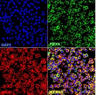
 Immunocytochemistry/ Immunofluorescence - Anti-Peroxiredoxin 6 antibody (ab59543) This image is courtesy of an Abreview submitted by Mahesh Shivananjappa
ab59543 staining Peroxiredoxin 6 in RAW264.7 cells from Mouse macrophages by ICC/IF (Immunocytochemistry/immunofluorescence). Cells were fixed with paraformaldehyde, permeabilized with Triton 0.1% + 2% BSA in PBS and blocked with 2% BSA for 60 minutes at 24°C. Samples were incubated with primary antibody (1/50 in PBS + 2% BSA) for 16 hours at 4°C. An Alexa Fluor®488-conjugated Goat anti-rabbit polyclonal(1/500) was used as the secondary antibody.
-
 Immunohistochemistry (Formalin/PFA-fixed paraffin-embedded sections) - Anti-Peroxiredoxin 6 antibody (ab59543)ab59543 at 1/100 dilution staining Peroxiredoxin 6 in astrocytes in Parkinson's disease by Immunohistochemistry. Secondary antibody anti-rabbit IgG conjugated to Cy3 (1/100).
 -
 Immunohistochemistry (Formalin/PFA-fixed paraffin-embedded sections) - Anti-Peroxiredoxin 6 antibody (ab59543)ab59543 at 1/1000 dilution staining Peroxiredoxin 6 in astrocytes in Parkinson's disease by IHC-P. Secondary antibody Donkey anti-rabbit conjugated to biotin.
 -
ICC/IF image of ab59543 stained HeLa cells. The cells were 100% methanol fixed (5 min) and then incubated in 1%BSA / 10% normal goat serum / 0.3M glycine in 0.1% PBS-Tween for 1h to permeabilise the cells and block non-specific protein-protein interactions. The cells were then incubated with the antibody (ab59543, 1/1000 dilution) overnight at +4°C. The secondary antibody (green) was Alexa Fluor® 488 goat anti-rabbit IgG (H+L) used at a 1/1000 dilution for 1h. Alexa Fluor® 594 WGA was used to label plasma membranes (red) at a 1/200 dilution for 1h. DAPI was used to stain the cell nuclei (blue) at a concentration of 1.43µM.


