Anti-Menin antibody - ChIP Grade (ab31902)
Key features and details
- Rabbit polyclonal to Menin - ChIP Grade
- Suitable for: IP, WB, ChIP, ICC/IF
- Knockout validated
- Reacts with: Mouse, Rat, Human
- Isotype: IgG
Overview
-
Product name
Anti-Menin antibody - ChIP Grade
See all Menin primary antibodies -
Description
Rabbit polyclonal to Menin - ChIP Grade -
Host species
Rabbit -
Tested Applications & Species
See all applications and species dataApplication Species ChIP HumanICC/IF HumanIP HumanWB MouseHuman -
Immunogen
Synthetic peptide conjugated to KLH derived from within residues 600 to the C-terminus of Human Menin.
Read Abcam's proprietary immunogen policy (Peptide available as ab32961.) -
Positive control
- Recombinant Menin protein (ab114387) can be used as a positive control in WB. This antibody gave a positive signal in the following Whole Cell Lysates: HeLa Jurkat A431 HEK 293 MEF1 (Mouse embryonic fibroblast cell line) PC12 (Rat adrenal pheochromocytoma cell line)
Images
-
Lane 1: Wild-type HAP1 cell lysate (20 µg)
Lane 2: Menin knockout HAP1 cell lysate (20 µg)
Lane 3: Jurkat cell lysate (20 µg)
Lane 4: A431 cell lysate (20 µg)
Lanes 1 - 4: Merged signal (red and green). Green - ab31902 observed at 74 kDa. Red - loading control, ab8245, observed at 37 kDa.ab31902 was shown to recognize Menin when Menin knockout samples were used, along with additional cross-reactive bands. Wild-type and Menin knockout samples were subjected to SDS-PAGE. ab31902 and ab8245 (loading control to GAPDH) were diluted 1 µg/mL and 1/2000 respectively and incubated overnight at 4°C. Blots were developed with Goat anti-Rabbit IgG H&L (IRDye® 800CW) preadsorbed (ab216773) and Goat anti-Mouse IgG H&L (IRDye® 680RD) preadsorbed (ab216776) secondary antibodies at 1/10 000 dilution for 1 h at room temperature before imaging.
-
All lanes : Anti-Menin antibody - ChIP Grade (ab31902) at 1 µg/ml
Lane 1 : HeLa (Human epithelial carcinoma cell line) Whole Cell Lysate
Lane 2 :Jurkat whole cell lysate (ab7899)
Lane 3 :A-431 whole cell lysate (ab7909)
Lane 4 :HEK-293 whole cell lysate (ab7902)
Lysates/proteins at 10 µg per lane.
Secondary
All lanes : IRDye 680 Conjugated Goat Anti-Rabbit IgG (H+L) at 1/15000 dilution
Performed under reducing conditions.
Predicted band size: 68 kDa
Observed band size: 75 kDa why is the actual band size different from the predicted?
Additional bands at: 31 kDa. We are unsure as to the identity of these extra bands. -
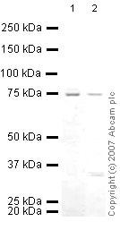
All lanes : Anti-Menin antibody - ChIP Grade (ab31902) at 1/250 dilution
Lane 1 : MEF1 (Mouse embryonic fibroblast cell line) Whole Cell Lysate
Lane 2 : PC12 (Rat adrenal pheochromocytoma cell line) Whole Cell Lysate
Lysates/proteins at 10 µg per lane.
Secondary
All lanes : IRDye 680 Conjugated Goat Anti-Rabbit IgG (H+L) at 1/10000 dilution
Performed under reducing conditions.
Predicted band size: 68 kDa
Observed band size: 75 kDa why is the actual band size different from the predicted?
-
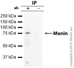
Menin was immunoprecipitated using 0.5mg Hela whole cell extract, 5µg of Rabbit polyclonal to Menin and 50µl of protein G magnetic beads (+). No antibody was added to the control (-).
The antibody was incubated under agitation with Protein G beads for 10min, Hela whole cell extract lysate diluted in RIPA buffer was added to each sample and incubated for a further 10min under agitation.
Proteins were eluted by addition of 40µl SDS loading buffer and incubated for 10min at 70oC; 10µl of each sample was separated on a SDS PAGE gel, transferred to a nitrocellulose membrane, blocked with 5% BSA and probed with ab31902.
Secondary: Mouse monoclonal [SB62a] Secondary Antibody to Rabbit IgG light chain (HRP) (ab99697).
Band: 75kDa: Menin. -
ChIP analysis of Menin along the PCNA promoter. High enrichment at PCNA PRO-2 and weak enrichment at PCNA PRO-1 is observed as previously described in literature.
Chromatin was prepared from HeLa cells according to the Abcam X-ChIP protocol. Cells were fixed with formaldehyde for 10 minutes. The ChIP was performed with 25µg of chromatin, 5µg of ab31902 (blue), and 20µl of Protein A/G sepharose beads. No antibody was added to the beads control (yellow). The immunoprecipitated DNA was quantified by real time PCR (Sybr green approach). Primers are located in the first kb of the transcribed region.
-
ICC/IF image of ab31902 stained human HEK 293 cells. The cells were PFA fixed (10 min), permabilised in TBS-T (20 min) and incubated with the antibody (ab31902, 1µg/ml) for 1h at room temperature. 1%BSA / 10% normal serum / 0.3M glycine was used to quench autofluorescence and block non-specific protein-protein interactions. The secondary antibody (green) was Alexa Fluor® 488 goat anti-rabbit IgG (H+L) used at a 1/1000 dilution for 1h. Alexa Fluor® 594 WGA was used to label plasma membranes (red). DAPI was used to stain the cell nuclei (blue).
-
ICC/IF image of ab31902 stained MCF7 cells. The cells were 4% PFA fixed (10 min) and then incubated in 1%BSA / 10% normal goat serum / 0.3M glycine in 0.1% PBS-Tween for 1h to permeabilise the cells and block non-specific protein-protein interactions. The cells were then incubated with the antibody (ab31902, 1µg/ml) overnight at +4°C. The secondary antibody (green) was Alexa Fluor® 488 goat anti-rabbit IgG (H+L) used at a 1/1000 dilution for 1h. Alexa Fluor® 594 WGA was used to label plasma membranes (red) at a 1/200 dilution for 1h. DAPI was used to stain the cell nuclei (blue) at a concentration of 1.43µM.


