Anti-LYVE1 antibody (ab33682)
")
Key features and details
- Rabbit polyclonal to LYVE1
- Suitable for: IHC, WB, ICC/IF
- Reacts with: Mouse, Human
- Isotype: IgG
Overview
-
Product name
Anti-LYVE1 antibody
See all LYVE1 primary antibodies -
Description
Rabbit polyclonal to LYVE1 -
Host species
Rabbit -
Tested Applications & Species
See all applications and species dataApplication Species ICC HumanIHC HumanWB Human
-
Immunogen
Synthetic peptide conjugated to KLH derived from within residues 300 to the C-terminus of Mouse LYVE1.
Read Abcam's proprietary immunogen policy (Peptide available as ab34093.) -
General notes
The Life Science industry has been in the grips of a reproducibility crisis for a number of years. Abcam is leading the way in addressing the problem with our range of recombinant monoclonal antibodies and knockout edited cell lines for gold-standard validation.
One factor contributing to the crisis is the use of antibodies that are not suitable. This can lead to misleading results and the use of incorrect data informing project assumptions and direction. To help address this challenge, we have introduced an application and species grid on our primary antibody datasheets to make it easy to simplify identification of the right antibody for your needs.
Learn more here.
Images
-
Immunocytochemistry - Anti-LYVE1 antibody (ab33682)
ab33682 staining LYVE1 in Jeg3 cells. The cells were fixed with 100% methanol (5 min), permeabilized with 0.1% PBS-Tween for 5 minutes and then blocked with 1% BSA/10% normal goat serum/0.3M glycine in 0.1%PBS-Tween for 1h. The cells were then incubated overnight at 4°C with ab33682 at 5µg/ml and ab7291, Mouse monoclonal [DM1A] to alpha Tubulin - Loading Control. Cells were then incubated with ab150081, Goat polyclonal Secondary Antibody to Rabbit IgG - H&L (Alexa Fluor® 488), pre-adsorbed at 1/1000 dilution (shown in green) and ab150120, Goat polyclonal Secondary Antibody to Mouse IgG - H&L (Alexa Fluor® 594), pre-adsorbed at 1/1000 dilution (shown in pseudocolour red). Nuclear DNA was labelled with DAPI (shown in blue).
Image was acquired with a high-content analyser (Operetta CLS, Perkin Elmer) and a maximum intensity projection of confocal sections is shown.
-
 Immunohistochemistry - Anti-LYVE1 antibody (ab33682)
IHC image of LYVE1 staining in Human Normal Colon formalin fixed paraffin embedded tissue section*, performed on a Leica Bond™ system using the standard protocol F. The section was pre-treated using heat mediated antigen retrieval with sodium citrate buffer (pH6, epitope retrieval solution 1) for 20 mins. The section was then incubated with ab33682, 0.1µg/ml, for 15 mins at room temperature and detected using an HRP conjugated compact polymer system. DAB was used as the chromogen. The section was then counterstained with haematoxylin and mounted with DPX.
For other IHC staining systems (automated and non-automated) customers should optimize variable parameters such as antigen retrieval conditions, primary antibody concentration and antibody incubation times.
*Tissue obtained from the Human Research Tissue Bank, supported by the NIHR Cambridge Biomedical Research Centre
-
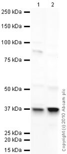 Western blot - Anti-LYVE1 antibody (ab33682)All lanes : Anti-LYVE1 antibody (ab33682) at 1 µg/ml
Lane 1 : A549 (Human lung adenocarcinoma epithelial cell line) Whole Cell Lysate
Lane 2 : Testis (Mouse) Tissue Lysate
Lysates/proteins at 10 µg per lane.
Secondary
All lanes : Goat polyclonal to Rabbit IgG - H&L - Pre-Adsorbed (HRP) at 1/3000 dilution
Developed using the ECL technique.
Performed under reducing conditions.
Predicted band size: 35 kDa
Observed band size: 37 kDa why is the actual band size different from the predicted?
Exposure time: 3 minutes
-
 Western blot - Anti-LYVE1 antibody (ab33682)All lanes : Anti-LYVE1 antibody (ab33682) at 1 µg/ml
Lane 1 : A549 (Human lung adenocarcinoma epithelial cell line) Whole Cell Lysate
Lane 2 : Testis (Mouse) Tissue Lysate
Lysates/proteins at 10 µg per lane.
Secondary
All lanes : Goat Anti-Rabbit IgG H&L (HRP) (ab97051) at 1/10000 dilution
Developed using the ECL technique.
Performed under reducing conditions.
Predicted band size: 35 kDa
Additional bands at: 35 kDa (possible mature (processed) protein), 42 kDa, 80 kDa. We are unsure as to the identity of these extra bands.
Exposure time: 20 minutesThe LYVE-1 protein has a predicted molecular weight of 35 kDa. The first 23 amino acids of the LYVE-1 sequence act as a signal sequence, and when cleaved the protein has an expected molecular weight of 32 kDa. We believe that the band observed at 35 kDa represents the 32 kDa product.
-
 Immunohistochemistry (Formalin/PFA-fixed paraffin-embedded sections) - Anti-LYVE1 antibody (ab33682) This image is courtesy of an anonymous Abreview
ab33682 staining LYVE1 in pig skin tissue sections by Immunohistochemistry (IHC-P - Formalin/PFA-fixed, paraffin-embedded sections). Tissue was fixed with formaldehyde and blocked with 3% serum for 30 minutes at 20°C; antigen retrieval was by heat mediation in citrate buffer, pH 6.0. Samples were incubated with primary antibody (1/200 in PBS) for 12 hours at 20°C. A HRP-conjugated goat anti-rabbit IgG polyclonal (1/200) was used as the secondary antibody.


