Anti-LYVE1 antibody (ab10278)
Key features and details
- Rabbit polyclonal to LYVE1
- Suitable for: IHC-Fr, Flow Cyt, IHC-P, WB
- Reacts with: Human, Recombinant fragment
- Isotype: IgG
Overview
-
Product name
Anti-LYVE1 antibody
See all LYVE1 primary antibodies -
Description
Rabbit polyclonal to LYVE1 -
Host species
Rabbit -
Tested applications
Suitable for: IHC-Fr, Flow Cyt, IHC-P, WBmore details -
Species reactivity
Reacts with: Human, Recombinant fragment
Predicted to work with: Rat
-
Immunogen
-
Positive control
- human colon carcinoma
Properties
-
Form
Liquid -
Storage instructions
Shipped at 4°C. Store at +4°C short term (1-2 weeks). Upon delivery aliquot. Store at -20°C. Avoid freeze / thaw cycle. -
Storage buffer
Constituent: PBS -
 Concentration information loading...
 -
Purity
Protein A purified -
Purification notes
Protein-A Chromatography (+his tag depleted). -
Primary antibody notes
The lymphatic vasculature forms a second circulatory system that drains extracellular fluid from the tissues and provides an exclusive environment in which immune cells can encounter and respond to foreign antigen. Recently a number of interesting molecules have been identified that may be exploited as markers for lymphatic endothelium, including the hyaluronan receptor LYVE1, PALE, VEGFR3, podoplanin. -
Clonality
Polyclonal -
Isotype
IgG -
Research areas
Images
-
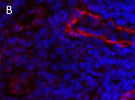
Immunohistochemistry (Frozen sections) analysis of human colon carcinoma tissue sections labelling LYVE1 with ab10278.
-
 Immunohistochemistry (Formalin/PFA-fixed paraffin-embedded sections) - Anti-LYVE1 antibody (ab10278)ab10278 (2µg/ml) staining LYVE1 in human colon using an automated system (DAKO Autostainer Plus). Using this protocol there is lymphatic endothelium staining of lymphatic ducts where blood vessel endothelium and smooth muscle is wholly negative.
Sections were rehydrated and antigen retrieved with the Dako 3-in-1 AR buffer citrate pH 6.0 in a DAKO PT Link. Slides were peroxidase blocked in 3% H2O2 in methanol for 10 minutes. They were then blocked with Dako Protein block for 10 minutes (containing casein 0.25% in PBS) then incubated with primary antibody for 20 minutes and detected with Dako Envision Flex amplification kit for 30 minutes. Colorimetric detection was completed with Diaminobenzidine for 5 minutes. Slides were counterstained with Haematoxylin and coverslipped under DePeX. Please note that, for manual staining, optimization of primary antibody concentration and incubation time is recommended. Signal amplification may be required.
-
All lanes : Anti-LYVE1 antibody (ab10278) at 1 µg/ml
Lane 1 : A549 (Human lung adenocarcinoma epithelial cell line) Whole Cell Lysate
Lane 4 : HepG2 (Human hepatocellular liver carcinoma cell line) Whole Cell Lysate
Lysates/proteins at 10 µg per lane.
Secondary
All lanes : Goat polyclonal to Rabbit IgG - H&L - Pre-Adsorbed (HRP) at 1/3000 dilution
Developed using the ECL technique.
Performed under reducing conditions.
Predicted band size: 35 kDa
Observed band size: 37 kDa why is the actual band size different from the predicted?
Additional bands at: 22 kDa, 55 kDa. We are unsure as to the identity of these extra bands.
Exposure time: 4 minutes
LYVE-1 contains a number of potential glycosylation sites (SwissProt) which may explain its migration at a higher molecular weight than predicted. -
Flow Cytometry analysis of human dermal microvascular endothelial cells (HDMVEC) labelling LYVE1 with ab10278.
-
 Immunohistochemistry (Frozen sections) - Anti-LYVE1 antibody (ab10278) This image is courtesy of an Abreview submitted by Dr Vyacheslav Ogay
Rat skin was fixed with paraformaldehyde in 15% saturated picric acid solution for 4hr. Prior to sectioning, the specimen was infiltrated in O.C.T. and frozen in isopentane. The frozen specimen was sectioned these were rinsed in PBS for 15 min to remove O.C.T. and incubated in a 3% sodium deoxycholate solution. The specimens were rinsed twice with distilled water and then with PBS three times. The sections were incubated in 10% normal goat serum for 12 hr at 4°C, then for 12 hr with ab10278. After washing with PBS, the specimens were incubated with Alexa Fluor® 555-conjugated goat anti-rabbit IgG (H+L) (1:500), for 12 hr at 4°C. The cell nuclei were counterstained with YoYo-1. Images were obtained by using confocal microscope.


