Anti-Histone H3 (citrulline R2 + R8 + R17) antibody (ab5103)
Key features and details
- Rabbit polyclonal to Histone H3 (citrulline R2 + R8 + R17)
- Suitable for: PepArr, ICC, WB
- Reacts with: Mouse, Rat, Human
- Isotype: IgG
Overview
-
Product name
Anti-Histone H3 (citrulline R2 + R8 + R17) antibody
See all Histone H3 primary antibodies -
Description
Rabbit polyclonal to Histone H3 (citrulline R2 + R8 + R17) -
Host species
Rabbit -
Specificity
ab5103 detects a 17 kDa band in single lane Western Blot. Peptide inhibition in Western Blot hasn't been processed. Modification specificity is determined by Peptide Array. ab5103 binds strongly to Histone H3 citrulline 2 + 8 + 17 peptide. -
Tested Applications & Species
See all applications and species dataApplication Species ICC HumanWB MouseRatHuman -
Immunogen
-
Positive control
- WB: Mouse and rat brain tissue lysates. HL60 + DMSO, NIH/3T3 and PC12 whole cell lysates. ICC: MCF7 cells.
-
General notes
Immunizing Peptide (Blocking) - ab32876.
Images
-
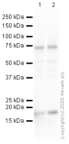
All lanes : Anti-Histone H3 (citrulline R2 + R8 + R17) antibody (ab5103) at 1 µg/ml
Lane 1 : Mouse brain tissue lysate
Lane 2 : Rat brain tissue lysate
Lysates/proteins at 10 µg per lane.
Secondary
All lanes : Goat polyclonal to Rabbit IgG - H&L - Pre-Adsorbed (HRP) at 1/50000 dilution
Predicted band size: 15 kDa
Observed band size: 17 kDa why is the actual band size different from the predicted?
Exposure time: 1 minuteBlocking buffer: 2% BSA
Gel type: MES
-
Immunocytochemistry analysis of ab5103 staining Histone H3 (citrulline R2 + R8 + R17) in MCF7 cells. The cells were fixed with 4% paraformaldehyde (10 min), permeabilized with 0.1% PBS-Triton X-100 for 5 minutes and then blocked with 1% BSA/10% normal goat serum/0.3M glycine in 0.1%PBS-Tween for 1h. The cells were then incubated overnight at 4°C with ab5103 at 1µg/mL and ab7291, Mouse monoclonal [DM1A] to alpha Tubulin - Loading Control. Cells were then incubated with ab150081, Goat polyclonal Secondary Antibody to Rabbit IgG - H&L (Alexa Fluor® 488), pre-adsorbed at 1/1000 dilution (shown in green) and ab150120, Goat polyclonal Secondary Antibody to Mouse IgG - H&L (Alexa Fluor® 594), pre-adsorbed at 1/1000 dilution (shown in pseudocolour red). Nuclear DNA was labelled with DAPI (shown in blue). Also suitable in cells fixed with 100% methanol (5 min).
-
All lanes : Anti-Histone H3 (citrulline R2 + R8 + R17) antibody (ab5103) at 1 µg/ml
Lane 1 : NIH/3T3 (Mouse embryo fibroblast cell line) nuclear lysate
Lane 2 : PC12 (Rat adrenal pheochromocytoma cell line) nuclear lysate
Lysates/proteins at 10 µg per lane.
Secondary
All lanes : Goat polyclonal to Rabbit IgG - H&L - Pre-Adsorbed (HRP) at 1/50000 dilution
Predicted band size: 15 kDa
Observed band size: 17 kDa why is the actual band size different from the predicted?
Exposure time: 1 minuteBlocking buffer: 2% BSA
Gel type: MES
-
All lanes : Anti-Histone H3 (citrulline R2 + R8 + R17) antibody (ab5103) at 0.2 µg/ml
Lane 1 : HL60 whole cell lysate (negative control)
Lane 2 : HL60 whole cell lysate + DMSO (solvent control)
Lane 3 : HL60 whole cell lysate + DMSO + Calcium Ionophore (positive control)
Lysates/proteins at 20 µg per lane.
Secondary
All lanes : Goat anti Rabbit IR680 at 1/10000 dilution
Performed under reducing conditions.
Predicted band size: 15 kDa
Observed band size: 17 kDa why is the actual band size different from the predicted?Loading Control: GAPDH
This blot was produced using a 4-12% Bis-tris gel under the MES buffer system. The gel was run at 200V for 35 minutes before being transferred onto a Nitrocellulose membrane at 30V for 70 minutes. The membrane was then blocked for an hour using Licor blocking buffer before being incubated with ab5103 overnight at 4°C. Antibody binding was detected using Goat anti Rabbit IR680 secondary at a 1:10,000 dilution for 1hr at room temperature and then imaged using the Licor Odyssey CLx.
-
All batches of ab5103 are tested in Peptide Array against peptides to different Histone H3 modifications. Six dilutions of each peptide are printed on to the Peptide Array in triplicate and results are averaged before being plotted on to a graph. Results show strong binding to Histone H3 - citrulline 2 + 8 + 17 peptide (ab32876), indicating that this antibody specifically recognises the Histone H3 - citrulline 2 + 8 + 17 modifications.
ab32876 - Histone H3 - citrulline 2 + 8 + 17
ab17566 - Histone H3 - unmodified
-
All lanes : Anti-Histone H3 (citrulline R2 + R8 + R17) antibody (ab5103) at 1 µg/ml
Lane 1 : HL60 (Human Caucasian promyelocytic leukaemia) DMSO and Calcium Ionophore treated Whole Cell Lysate with with 5% BSA
Lane 2 : HL60 (Human Caucasian promyelocytic leukaemia) DMSO and Calcium Ionophore treated Whole Cell Lysate with with 5% milk
Lane 3 : HL60 (Human Caucasian promyelocytic leukaemia) DMSO and Calcium Ionophore treated Whole Cell Lysate with with 3% milk
Lysates/proteins at 10 µg per lane.
Secondary
All lanes : Goat Anti-Rabbit IgG H&L (HRP) (ab97051) at 1/10000 dilution
Developed using the ECL technique.
Performed under reducing conditions.
Predicted band size: 15 kDa
Observed band size: 17 kDa why is the actual band size different from the predicted?
Exposure time: 30 secondsAbcam recommends using milk as the blocking agent. Abcam welcomes customer feedback and would appreciate any comments regarding this product and the data presented above .
Blots were developled with Goat Anti-Rabbit IgG H&L (HRP) (ab97051) secondary antibody


