Anti-Gephyrin antibody (ab32206)
")
Key features and details
- Rabbit polyclonal to Gephyrin
- Suitable for: ICC/IF, WB
- Reacts with: Mouse, Rat
- Isotype: IgG
Overview
-
Product name
Anti-Gephyrin antibody
See all Gephyrin primary antibodies -
Description
Rabbit polyclonal to Gephyrin -
Host species
Rabbit -
Tested applications
Suitable for: ICC/IF, WBmore details -
Species reactivity
Reacts with: Mouse, Rat
Predicted to work with: Chicken, Human, Xenopus laevis
-
Immunogen
-
Positive control
- WB: Mouse brain and liver tissue lysate, rat brain and liver tissue lysate. ICC/IF: PC-12 cells.
Properties
-
Form
Liquid -
Storage instructions
Shipped at 4°C. Store at +4°C short term (1-2 weeks). Upon delivery aliquot. Store at -20°C or -80°C. Avoid freeze / thaw cycle. -
Storage buffer
pH: 7.40
Preservative: 0.02% Sodium azide
Constituent: PBS
Batches of this product that have a concentration Concentration information loading...
Purity
Immunogen affinity purifiedClonality
PolyclonalIsotype
IgGResearch areas
Associated products
-
Compatible Secondaries
-
Isotype control
-
Positive Controls
-
Recombinant Protein
Applications
Our Abpromise guarantee covers the use of ab32206 in the following tested applications.
The application notes include recommended starting dilutions; optimal dilutions/concentrations should be determined by the end user.
Application Abreviews Notes ICC/IF Use a concentration of 1 µg/ml. WB Use a concentration of 1 µg/ml. Detects a band of approximately 93,75,54 kDa (predicted molecular weight: 83 kDa). The 93 kDa band corresponds to Isoform 1 of Gephyrin as observed in the literature. We suspect that the 75 kDa band corresponds to another isoform of Gephyrin. The identity of the 54 kDa band is unknown. All three of the bands we observe on WB are partially blocked by addition of the immunising peptide (ab32205).
Target
-
Function
Microtubule-associated protein involved in membrane protein-cytoskeleton interactions. It is thought to anchor the inhibitory glycine receptor (GLYR) to subsynaptic microtubules (By similarity). Catalyzes two steps in the biosynthesis of the molybdenum cofactor. In the first step, molybdopterin is adenylated. Subsequently, molybdate is inserted into adenylated molybdopterin and AMP is released. -
Pathway
Cofactor biosynthesis; molybdopterin biosynthesis. -
Involvement in disease
Defects in GPHN are the cause of molybdenum cofactor deficiency type C (MOCOD type C) [MIM:252150]. MOCOD type C is an autosomal recessive disease which leads to the pleiotropic loss of all molybdoenzyme activities and is characterized by severe neurological damage, neonatal seizures and early childhood death.
Defects in GPHN are a cause of startle disease (STHE) [MIM:149400]; also known as hyperekplexia. STHE is a genetically heterogeneous neurologic disorder characterized by muscular rigidity of central nervous system origin, particularly in the neonatal period, and by an exaggerated startle response to unexpected acoustic or tactile stimuli. -
Sequence similarities
In the N-terminal section; belongs to the moaB/mog family.
In the C-terminal section; belongs to the moeA family. -
Cellular localization
Cell junction > synapse. Cell junction > synapse > postsynaptic cell membrane. Cytoplasm > cytoskeleton. Cytoplasmic face of glycinergic postsynaptic membranes. - Information by UniProt
-
Database links
- Entrez Gene: 428878 Chicken
- Entrez Gene: 10243 Human
- Entrez Gene: 268566 Mouse
- Entrez Gene: 64845 Rat
- Entrez Gene: 779372 Xenopus laevis
- Omim: 603930 Human
- SwissProt: Q9PW38 Chicken
- SwissProt: Q9NQX3 Human
see all -
Alternative names
- Domain E antibody
- Domain G antibody
- GEPH antibody
see all
Images
-
Western blot - Anti-Gephyrin antibody (ab32206)All lanes : ab32206 at 1 µg/ml
Lane 1 : Mouse Brain Tissue Lysate
Lane 2 : Mouse Liver Tissue Lysate
Lane 3 : Rat Brain Tissue Lysate
Lane 4 : Rat Liver Tissue Lysate
Lysates/proteins at 10 µg per lane.
Secondary
All lanes : Goat polyclonal to Rabbit IgG (Alexa Fluor® 680) at 1/1000 dilution
Developed using the ECL technique.
Performed under reducing conditions.
Predicted band size: 83 kDa
Observed band size: 90 kDa why is the actual band size different from the predicted?
Exposure time: 20 minutesWe are unsure of the identity of the 89kDa band observed in brain lysates, however this was consistent across both species tested.
Abcam recommends using milk as the blocking agent. Abcam welcomes customer feedback and would appreciate any comments regarding this product and the data presented above.
-
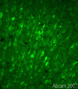 Immunohistochemistry (PFA perfusion fixed frozen sections) - Anti-Gephyrin antibody (ab32206)This image is courtesy of Sophie Pezet, CNRS, Paris, France
Immunofluorescent staining for Gephyrin in rat brain cortex using Rabbit polyclonal to Gephyrin (ab32206; 1/1000). The staining is located in the cytoplasm and in some of the processes of cortical neurons. This staining is observed in many other brain areas. The picture was acquired using a X20 objective. Protocol details: Rats were intracardially perfused with 4% paraformaldehyde. Whole brain tissue was post-fixed overnight in the same fixative, and cryoprotected in 20% sucrose and frozen in OCT. 30 µm coronal sections were cut by cyrostat for use in fre floating IHC. Primary antibody ab32206 was incubated overnight at 1/1000 at room temperature. Secondary antibody Alexa fluor® 488 1/1000 was incubated for 2 hours at room temperature.
-
 Immunocytochemistry/ Immunofluorescence - Anti-Gephyrin antibody (ab32206)
ICC/IF image of ab32206 stained PC-12 (Rat adrenal gland pheochromocytoma cell line) cells. The cells were fixed in 100% methanol for 5 minutes and then incubated in 1% BSA / 10% normal goat serum / 0.3M glycine in 0.1% PBS-Tween for 1 hour to permeabilize the cells and block non-specific protein-protein interactions. The cells were then incubated with the antibody (ab32206, 1 µg/ml) overnight at +4°C. The secondary antibody (green) was ab96899, DyLight® 488 goat anti-rabbit IgG (H+L) used at a 1/250 dilution for 1 hour. Alexa Fluor® 594 WGA was used to label plasma membranes (red) at a 1/200 dilution for 1 hour. DAPI was used to stain the cell nuclei (blue) at a concentration of 1.43 µM.
Protocols
References (13)
ab32206 has been referenced in 13 publications.
- Scaduto P et al. Preservation of global synaptic excitatory to inhibitory ratio during long postmortem intervals. Sci Rep 10:8626 (2020). PubMed: 32451470
- Wang S et al. Macroglia-derived thrombospondin 2 regulates alterations of presynaptic proteins of retinal neurons following elevated hydrostatic pressure. PLoS One 12:e0185388 (2017). PubMed: 28953973
- Notaras M et al. The BDNF Val66Met polymorphism regulates glucocorticoid-induced corticohippocampal remodeling and behavioral despair. Transl Psychiatry 7:e1233 (2017). WB ; Mouse . PubMed: 28926000
- Belmer A et al. Mapping the connectivity of serotonin transporter immunoreactive axons to excitatory and inhibitory neurochemical synapses in the mouse limbic brain. Brain Struct Funct 222:1297-1314 (2017). PubMed: 27485750
- Lu P et al. Prolonged human neural stem cell maturation supports recovery in injured rodent CNS. J Clin Invest 127:3287-3299 (2017). PubMed: 28825600
Images
-
Western blot - Anti-Gephyrin antibody (ab32206)All lanes : ab32206 at 1 µg/ml
Lane 1 : Mouse Brain Tissue Lysate
Lane 2 : Mouse Liver Tissue Lysate
Lane 3 : Rat Brain Tissue Lysate
Lane 4 : Rat Liver Tissue Lysate
Lysates/proteins at 10 µg per lane.
Secondary
All lanes : Goat polyclonal to Rabbit IgG (Alexa Fluor® 680) at 1/1000 dilution
Developed using the ECL technique.
Performed under reducing conditions.
Predicted band size: 83 kDa
Observed band size: 90 kDa why is the actual band size different from the predicted?
Exposure time: 20 minutesWe are unsure of the identity of the 89kDa band observed in brain lysates, however this was consistent across both species tested.
Abcam recommends using milk as the blocking agent. Abcam welcomes customer feedback and would appreciate any comments regarding this product and the data presented above.
-
Immunohistochemistry (PFA perfusion fixed frozen sections) - Anti-Gephyrin antibody (ab32206) This image is courtesy of Sophie Pezet, CNRS, Paris, France
Immunofluorescent staining for Gephyrin in rat brain cortex using Rabbit polyclonal to Gephyrin (ab32206; 1/1000). The staining is located in the cytoplasm and in some of the processes of cortical neurons. This staining is observed in many other brain areas. The picture was acquired using a X20 objective. Protocol details: Rats were intracardially perfused with 4% paraformaldehyde. Whole brain tissue was post-fixed overnight in the same fixative, and cryoprotected in 20% sucrose and frozen in OCT. 30 µm coronal sections were cut by cyrostat for use in fre floating IHC. Primary antibody ab32206 was incubated overnight at 1/1000 at room temperature. Secondary antibody Alexa fluor® 488 1/1000 was incubated for 2 hours at room temperature.
-
Immunocytochemistry/ Immunofluorescence - Anti-Gephyrin antibody (ab32206)
ICC/IF image of ab32206 stained PC-12 (Rat adrenal gland pheochromocytoma cell line) cells. The cells were fixed in 100% methanol for 5 minutes and then incubated in 1% BSA / 10% normal goat serum / 0.3M glycine in 0.1% PBS-Tween for 1 hour to permeabilize the cells and block non-specific protein-protein interactions. The cells were then incubated with the antibody (ab32206, 1 µg/ml) overnight at +4°C. The secondary antibody (green) was ab96899, DyLight® 488 goat anti-rabbit IgG (H+L) used at a 1/250 dilution for 1 hour. Alexa Fluor® 594 WGA was used to label plasma membranes (red) at a 1/200 dilution for 1 hour. DAPI was used to stain the cell nuclei (blue) at a concentration of 1.43 µM.


