Anti-DNA PKcs (phospho S2056) antibody (ab18192)
 antibody (ab18192)")
Key features and details
- Rabbit polyclonal to DNA PKcs (phospho S2056)
- Suitable for: WB, ELISA, ICC/IF
- Reacts with: Human
- Isotype: IgG
Overview
-
Product name
Anti-DNA PKcs (phospho S2056) antibody
See all DNA PKcs primary antibodies -
Description
Rabbit polyclonal to DNA PKcs (phospho S2056) -
Host species
Rabbit -
Specificity
This antibody specifically recognizes a band at ~460 kDa in HeLa cells that have been treated with ionizing radiation, that is not detected in untreated cells. This band can also be competed away by the immunizing modified peptide, but not the unmodified peptide containing the same amino acid sequence. All batches of this antibody are screened in ELISA and show high titres against the immunising peptide. Reactivity with the unmodified DNA PKcs peptide is minimal. We now predict that this antibody will cross react with mouse DNA PKcs, as the mouse sequence has been more extensively reviewed on uniprot (P97313), now indicating that mouse DNA PKcs S2053, which corresponds to human S2056, is also phosphorylated. We welcome any feedback from researchers who have used this antibody with mouse samples. -
Tested Applications & Species
See all applications and species dataApplication Species ICC/IF HumanWB Human
-
Immunogen
Synthetic peptide conjugated to KLH derived from within residues 2050 - 2150 of Human DNA PKcs.
Read Abcam's proprietary immunogen policy (Peptide available as ab20406.) -
General notes
The Life Science industry has been in the grips of a reproducibility crisis for a number of years. Abcam is leading the way in addressing the problem with our range of recombinant monoclonal antibodies and knockout edited cell lines for gold-standard validation.
One factor contributing to the crisis is the use of antibodies that are not suitable. This can lead to misleading results and the use of incorrect data informing project assumptions and direction. To help address this challenge, we have introduced an application and species grid on our primary antibody datasheets to make it easy to simplify identification of the right antibody for your needs.
Learn more here.
Images
-
Western blot - Anti-DNA PKcs (phospho S2056) antibody (ab18192)
Lane 1: Wild type HAP1 whole cell lysate (20 µg)
Lane 2: HAP1 Parental Camptothecin (1um 1hr) whole cell lysate (20 µg)
Lane 3: HAP1 PRKDC KO Untreated whole cell lysate (20 µg)
Lane 4: HAP1 PRKDC KO camptothecin (1um 1hr) whole cell lysate (20ug)Lane 5: SHSY-5Y untreated whole cell lysate (20ug)
Lane 6: SHSY-5Y Camptothecin treated (1uM, 1hr) whole tissue lysate (20 µg)
Lanes 1 - 4: Merged signal (red and green). Green - ab18192 observed at 450 kDa. Red - loading control, ab6301, observed at 85 kDa.
ab18192 was shown to specifically react with DNA PKcs (phospho S2056) in wild-type HAP1 cells along with additional cross reactive bands. Uplift was also seen with Camptothecin treatment and no bands were observed when DNA PKcs (phospho S2056) knockout cellsd. Wild-type and DNA PKcs (phospho S2056) knockout samples were subjected to SDS-PAGE. ab18192 and ab6301 (Mouse anti-Beta Catenin loading control) were incubated overnight at 4°C at 1 µg/ml and 1/10,000 dilution respectively. Blots were developed with Goat anti-Rabbit IgG H&L (IRDye® 800CW) preabsorbed (ab216773) and Goat anti-Mouse IgG H&L (IRDye® 680RD) preabsorbed (ab216776) secondary antibodies at 1/10,000 dilution for 1 hour at room temperature before imaging.
-
 Immunocytochemistry/ Immunofluorescence - Anti-DNA PKcs (phospho S2056) antibody (ab18192)
ICC/IF image of ab18192 stained UV treated HeLa cells. The cells were 100% methanol fixed (5 min) then permeabilised using 0.1% PBS-Triton and then incubated in 1%BSA / 10% normal goat serum / 0.3M glycine in 0.1% PBS-Tween for 1h to further permeabilise the cells and block non-specific protein-protein interactions. The cells were then incubated with the antibody ab18192 at 5µg/ml overnight at +4°C. The secondary antibody (pseudo-colored green) was Alexa Fluor® 488 goat anti- rabbit (ab150081) IgG (H+L) preadsorbed, used at a 1/1000 dilution for 1h. Alexa Fluor® 594 WGA was used to label plasma membranes (pseudo-colored red) at a 1/200 dilution for 1h at room temperature. DAPI was used to stain the cell nuclei (pseudo-colored blue) at a concentration of 1.43µM for 1hour at room temperature.
-
 Immunocytochemistry/ Immunofluorescence - Anti-DNA PKcs (phospho S2056) antibody (ab18192) Furusawa Y et al. DNA double-strand breaks induced by cavitational mechanical effects of ultrasound in cancer cell lines. PLoS One 7:e29012 (2012). Reproduced under the Creative Commons license http://creativecommons.org/licenses/by/4.0/
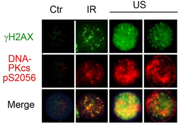
U937 (Human histiocytic lymphoma cell line) cells were paraformaldehyde-fixe. Control and treated cells were permeabilized/blocked with 2% BSA/0.05% Triton X-100/Tris-buffered saline, and immunostained for DNA PKcs (phospho S2056) (Red) for 2 h with ab18192 at 1/600 dilution.
Green staining shows γH2AX. IR = Ionzing radiation. US = Ultrasound.
-
 Western blot - Anti-DNA PKcs (phospho S2056) antibody (ab18192)All lanes : Anti-DNA PKcs (phospho S2056) antibody (ab18192) at 1 µg/ml
Lane 1 : HeLa Gamma Irradiated Whole Cell Lysate at 20 µg
Lane 2 : 20ug of untreated HeLa cell extract
Lane 3 : HeLa Gamma Irradiated Whole Cell Lysate at 20 µg with Human DNA PKcs (phospho S2056) peptide (ab20406) at 1 µg/ml
Lane 4 : 20ug of untreated HeLa cell extract with Human DNA PKcs (phospho S2056) peptide (ab20406) at 1 µg/ml
Lane 5 : HeLa Gamma Irradiated Whole Cell Lysate at 20 µg withHuman DNA PKcs peptide (ab20407) at 1 µg/ml
Lane 6 : 20ug of untreated HeLa cell extract withHuman DNA PKcs peptide (ab20407) at 1 µg/ml
Secondary
Lanes 1 & 3 & 5 : Alexa Fluor Goat polyclonal to Rabbit IgG (700) at 1/10000 dilution
Lanes 2 & 4 & 6 : Alexa Fluor Goat polyclonal to Rabbit IgG (700)
at 1/10000 dilution
Developed using the ECL technique.
Performed under reducing conditions.
Predicted band size: 460 kDa
Observed band size: 460 kDa
Additional bands at: 270 kDa (possible cleavage fragment), 270 kDa (possible cross reactivity)ab18192 specifically recognizes a band at ~460 kDa corresponding to DNA PKcs in HeLa cells that have been treated with ionizing radiation (lane 1). This band is not detected in untreated cells (lane 2). The activity of the antibody is quenched by the addition of the immunizing (modified) peptide, ab20406 (lanes 3) but not the unmodified peptide, ab20407 (lane 5).
For the ab13823 irradiated HeLa cell lysate, the 4 hour post-treatment extract was used.
-
 ELISA - Anti-DNA PKcs (phospho S2056) antibody (ab18192)
Serially diluted ab18192 was bound to immobilised DNA PKcs phospho peptide (2052 - 2062; P-S2056) or DNA PKcs control peptide (2052 - 2062; both at 1 microgram x mL-1). The antibody was detected by HRP-labelled goat anti-rabbit IgG (ab97080; diluted 50000 times) and signal was developed with TMB substrate.
-
 Immunocytochemistry/ Immunofluorescence - Anti-DNA PKcs (phospho S2056) antibody (ab18192) Image from Furusawa Y et al., PLoS One. 2012;7(1):e29012. Epub 2012 Jan 3. Fig 2.; doi:10.1371/journal.pone.0029012; January 3, 2012, PLoS ONE 7(1): e29012.ab18192 staining DNA PKcs (phospho S2056) in leukaemia cell lines by Immunocytochemistry/ Immunofluorescence.
Cells were either untreated (control) or treated with ionizing radiation (IR) or ultrasound (US). Following treatment, cells were fixed with paraformaldehyde and permabilized/blocked with 2% BSA/0.05% Triton X-100/Tris-buffered saline. Samples were incubated with primary antibody (1/600 in diluent) for 2 hours. An AlexaFluor®555-conjugated anti-rabbit IgG (1/400) was used as the secondary antibody. -
 Western blot - Anti-DNA PKcs (phospho S2056) antibody (ab18192)All lanes : Anti-DNA PKcs (phospho S2056) antibody (ab18192) at 1 µg/ml
Lane 1 : HeLa (Human epithelial carcinoma cell line) Whole Cell Lysate
Lane 2 : Hela Whole Cell Lysate - Bleomycin Treated (20U/ml)
Lane 3 : HeLa (Human epithelial carcinoma cell line) Whole Cell Lysate with Human DNA PKcs (phospho S2056) peptide (ab20406) at 1 µg/ml
Lane 4 : Hela Whole Cell Lysate - Bleomycin Treated (20U/ml) with Human DNA PKcs (phospho S2056) peptide (ab20406) at 1 µg/ml
Lane 5 : HeLa (Human epithelial carcinoma cell line) Whole Cell Lysate withHuman DNA PKcs peptide (ab20407) at 1 µg/ml
Lane 6 : Hela Whole Cell Lysate - Bleomycin Treated (20U/ml) withHuman DNA PKcs peptide (ab20407) at 1 µg/ml
Lysates/proteins at 20 µg per lane.
Secondary
All lanes : Goat polyclonal to Rabbit IgG - H&L - Pre-Adsorbed (HRP) at 1/3000 dilution
Performed under reducing conditions.
Predicted band size: 460 kDa
Observed band size: 460 kDa
Additional bands at: 117 kDa, 150 kDa, 270 kDa, 50 kDa. We are unsure as to the identity of these extra bands.
Exposure time: 15 minutes


