Anti-CPS1 antibody (ab45956)
")
Key features and details
- Rabbit polyclonal to CPS1
- Suitable for: WB, ICC/IF, IHC-P
- Reacts with: Human
- Isotype: IgG
Overview
-
Product name
Anti-CPS1 antibody
See all CPS1 primary antibodies -
Description
Rabbit polyclonal to CPS1 -
Host species
Rabbit -
Tested applications
Suitable for: WB, ICC/IF, IHC-Pmore details -
Species reactivity
Reacts with: Human
Predicted to work with: Mouse, Rat
-
Immunogen
Synthetic peptide corresponding to Human CPS1 aa 800-900 conjugated to keyhole limpet haemocyanin.
(Peptide available asab45955) -
General notes
The Life Science industry has been in the grips of a reproducibility crisis for a number of years. Abcam is leading the way in addressing this with our range of recombinant monoclonal antibodies and knockout edited cell lines for gold-standard validation. Please check that this product meets your needs before purchasing.
If you have any questions, special requirements or concerns, please send us an inquiry and/or contact our Support team ahead of purchase. Recommended alternatives for this product can be found below, along with publications, customer reviews and Q&As
Properties
-
Form
Liquid -
Storage instructions
Shipped at 4°C. Store at +4°C short term (1-2 weeks). Upon delivery aliquot. Store at -20°C or -80°C. Avoid freeze / thaw cycle. -
Storage buffer
pH: 7.40
Preservative: 0.02% Sodium azide
Constituent: PBS
Batches of this product that have a concentration Concentration information loading...
Purity
Immunogen affinity purifiedClonality
PolyclonalIsotype
IgGResearch areas
Associated products
-
Compatible Secondaries
-
Isotype control
-
Recombinant Protein
Applications
The Abpromise guarantee
Our Abpromise guarantee covers the use of ab45956 in the following tested applications.
The application notes include recommended starting dilutions; optimal dilutions/concentrations should be determined by the end user.
Application Abreviews Notes WB (2) Use a concentration of 1 µg/ml. Detects a band of approximately 150 kDa (predicted molecular weight: 140 kDa).ICC/IF (1) Use a concentration of 1 µg/ml.IHC-P Use a concentration of 1 µg/ml. Perform heat mediated antigen retrieval before commencing with IHC staining protocol.Notes WB
Use a concentration of 1 µg/ml. Detects a band of approximately 150 kDa (predicted molecular weight: 140 kDa).ICC/IF
Use a concentration of 1 µg/ml.IHC-P
Use a concentration of 1 µg/ml. Perform heat mediated antigen retrieval before commencing with IHC staining protocol.Target
-
Function
Involved in the urea cycle of ureotelic animals where the enzyme plays an important role in removing excess ammonia from the cell. -
Tissue specificity
Primarily in the liver and small intestine. -
Involvement in disease
Defects in CPS1 are the cause of carbamoyl phosphate synthetase 1 deficiency (CPS1D) [MIM:237300]. CPS1D is an autosomal recessive disorder of the urea cycle causing hyperammonemia. Clinical features include protein intolerance, intermittent ataxia, seizures, lethargy, developmental delay and mental retardation.
Note=Genetic variations in CPS1 influence the availability of precursors for nitric oxide (NO) synthesis and play a role in clinical situations where endogenous NO production is critically important, such as neonatal pulmonary hypertension, increased pulmonary artery pressure following surgical repair of congenital heart defects or hepatovenocclusive disease following bone marrow transplantation. Infants with neonatal pulmonary hypertension homozygous for Thr-1406 have lower L-arginine concentrations than neonates homozygous for Asn-1406. -
Sequence similarities
Contains 2 ATP-grasp domains.
Contains 1 glutamine amidotransferase type-1 domain. -
Domain
The type-1 glutamine amidotransferase domain is defective. -
Cellular localization
Mitochondrion. - Information by UniProt
-
Database links
- Entrez Gene: 1373 Human
- Entrez Gene: 227231 Mouse
- Entrez Gene: 497840 Rat
- Omim: 608307 Human
- SwissProt: P31327 Human
- SwissProt: Q8C196 Mouse
- SwissProt: P07756 Rat
- Unigene: 149252 Human
see all -
Alternative names
- Carbamoyl phosphate synthase [ammonia] antibody
- Carbamoyl phosphate synthase [ammonia] mitochondrial antibody
- Carbamoyl phosphate synthase antibody
see all
Images
-
Western blot - Anti-CPS1 antibody (ab45956)All lanes : Anti-CPS1 antibody (ab45956) at 1 µg/ml
Lane 1 : HeLa (Human epithelial carcinoma cell line) Whole Cell Lysate
Lane 2 : A431 (Human epithelial carcinoma cell line) Whole Cell Lysate
Lane 3 : HepG2 (Human hepatocellular liver carcinoma cell line) Whole Cell Lysate
Lysates/proteins at 10 µg per lane.
Secondary
All lanes : Goat polyclonal to Rabbit IgG - H&L - Pre-Adsorbed (HRP) at 1/3000 dilution
Performed under reducing conditions.
Predicted band size: 140 kDa
Observed band size: 150 kDa why is the actual band size different from the predicted? -
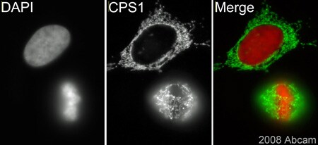 Immunocytochemistry/ Immunofluorescence - Anti-CPS1 antibody (ab45956)This image is courtesy of an Abreview submitted by Dr Kirk McManusab45956 (1/500) staining CPS1 in HeLa cells (green). Cells were fixed in methanol and counterstained with DAPI in order to highlight the nucleus (red). Please refer to abreview for further experimental details.
 -
 Immunocytochemistry/ Immunofluorescence - Anti-CPS1 antibody (ab45956)ICC/IF image of ab45956 stained human HeLa cells. The cells were PFA fixed (10 min), permabilised in PBS-T (20 min) and incubated with the antibody (ab45956, 1µg/ml) for 1h at room temperature. 1%BSA / 10% normal goat serum / 0.3M glycine was used to quench autofluorescence and block non-specific protein-protein interactions. The secondary antibody (green) was Alexa Fluor® 488 goat anti-rabbit IgG (H+L) used at a 1/1000 dilution for 1h. Alexa Fluor® 594 WGA was used to label plasma membranes (red). DAPI was used to stain the cell nuclei (blue).
 -
 Immunohistochemistry (Formalin/PFA-fixed paraffin-embedded sections) - Anti-CPS1 antibody (ab45956)IHC image of CPS1 staining in human liver carcinoma FFPE section, performed on a BondTM system using the standard protocol F. The section was pre-treated using heat mediated antigen retrieval with sodium citrate buffer (pH6, epitope retrieval solution 1) for 20 mins. The section was then incubated with ab45956, 1µg/ml, for 8 mins at room temperature and detected using an HRP conjugated compact polymer system. DAB was used as the chromogen. The section was then counterstained with haematoxylin and mounted with DPX.
Protocols
Datasheets and documents
-
Datasheet download
References (13)
ab45956 has been referenced in 13 publications.
- Kallabis S et al. High-throughput proteomics fiber typing (ProFiT) for comprehensive characterization of single skeletal muscle fibers. Skelet Muscle 10:7 (2020). PubMed: 32293536
- Greene KS et al. SIRT5 stabilizes mitochondrial glutaminase and supports breast cancer tumorigenesis. Proc Natl Acad Sci U S A N/A:N/A (2019). PubMed: 31843902
- Li X et al. Probiotics Ameliorate Colon Epithelial Injury Induced by Ambient Ultrafine Particles Exposure. Adv Sci (Weinh) 6:1900972 (2019). PubMed: 31559135
- El-Sheikh RM et al. Carbamoyl phosphate synthetase 1 (CPS1) as a prognostic marker in chronic hepatitis C infection. APMIS 127:93-105 (2019). PubMed: 30698308
- Soria LR et al. Hepatic glutamine synthetase augmentation enhances ammonia detoxification. J Inherit Metab Dis 42:1128-1135 (2019). PubMed: 30724386
Images
-
Western blot - Anti-CPS1 antibody (ab45956)All lanes : Anti-CPS1 antibody (ab45956) at 1 µg/ml
Lane 1 : HeLa (Human epithelial carcinoma cell line) Whole Cell Lysate
Lane 2 : A431 (Human epithelial carcinoma cell line) Whole Cell Lysate
Lane 3 : HepG2 (Human hepatocellular liver carcinoma cell line) Whole Cell Lysate
Lysates/proteins at 10 µg per lane.
Secondary
All lanes : Goat polyclonal to Rabbit IgG - H&L - Pre-Adsorbed (HRP) at 1/3000 dilution
Performed under reducing conditions.
Predicted band size: 140 kDa
Observed band size: 150 kDa why is the actual band size different from the predicted?
-
Immunocytochemistry/ Immunofluorescence - Anti-CPS1 antibody (ab45956) This image is courtesy of an Abreview submitted by Dr Kirk McManusab45956 (1/500) staining CPS1 in HeLa cells (green). Cells were fixed in methanol and counterstained with DAPI in order to highlight the nucleus (red). Please refer to abreview for further experimental details.
-
Immunocytochemistry/ Immunofluorescence - Anti-CPS1 antibody (ab45956)ICC/IF image of ab45956 stained human HeLa cells. The cells were PFA fixed (10 min), permabilised in PBS-T (20 min) and incubated with the antibody (ab45956, 1µg/ml) for 1h at room temperature. 1%BSA / 10% normal goat serum / 0.3M glycine was used to quench autofluorescence and block non-specific protein-protein interactions. The secondary antibody (green) was Alexa Fluor® 488 goat anti-rabbit IgG (H+L) used at a 1/1000 dilution for 1h. Alexa Fluor® 594 WGA was used to label plasma membranes (red). DAPI was used to stain the cell nuclei (blue).
-
Immunohistochemistry (Formalin/PFA-fixed paraffin-embedded sections) - Anti-CPS1 antibody (ab45956)IHC image of CPS1 staining in human liver carcinoma FFPE section, performed on a BondTM system using the standard protocol F. The section was pre-treated using heat mediated antigen retrieval with sodium citrate buffer (pH6, epitope retrieval solution 1) for 20 mins. The section was then incubated with ab45956, 1µg/ml, for 8 mins at room temperature and detected using an HRP conjugated compact polymer system. DAB was used as the chromogen. The section was then counterstained with haematoxylin and mounted with DPX.


