Anti-Aurora A antibody - Centrosome Marker (ab1287)
Key features and details
- Rabbit polyclonal to Aurora A - Centrosome Marker
- Suitable for: IHC-P, WB, IP
- Reacts with: Human
- Isotype: IgG
Overview
-
Product name
Anti-Aurora A antibody - Centrosome Marker
See all Aurora A primary antibodies -
Description
Rabbit polyclonal to Aurora A - Centrosome Marker -
Host species
Rabbit -
Tested applications
Suitable for: IHC-P, WB, IPmore details -
Species reactivity
Reacts with: Human
Predicted to work with: Chimpanzee
-
Immunogen
Synthetic peptide (Human) conjugated to KLH - which represented a portion of human Serine/Threonine Kinase 15 encoded within exon 5 (LocusLink ID 8465)
Properties
-
Form
Liquid -
Storage instructions
Shipped at 4°C. Upon delivery aliquot and store at -20°C. Avoid freeze / thaw cycles. -
Storage buffer
pH: 7
Preservative: 0.1% Sodium azide
Constituents: 0.021% PBS, 1.764% Sodium citrate, 1.815% Tris -
 Concentration information loading...
 -
Purity
Immunogen affinity purified -
Clonality
Polyclonal -
Isotype
IgG -
Research areas
Images
-
Anti-Aurora A antibody - Centrosome Marker (ab1287) at 1 µg/ml + Human liver tissue lysate - total protein (ab29889) at 10 µg
Secondary
Goat polyclonal to Rabbit IgG - H&L - Pre-Adsorbed (HRP) at 1/3000 dilution
Predicted band size: 48 kDa
Observed band size: 48 kDa
Additional bands at: 20 kDa, 25 kDa, 28 kDa, 37 kDa. We are unsure as to the identity of these extra bands.
-
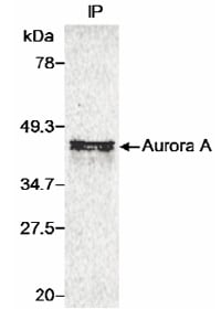
Immunoprecipitation of Human Aurora A.
35S-Met labeled whole cell lysate from cells transfected with a human Aurora A expression construct. ab1287 was used at 4 µg/ml for IP. Detection: Autoradiography. -
 Immunohistochemistry (Formalin/PFA-fixed paraffin-embedded sections) - Anti-Aurora A antibody - Centrosome Marker (ab1287)ab1287 (1/250) detecting Aurora A in paraffin embedded Human pancreatic tumour tissue sections. Detection: DAB staining;


