Biotin Anti-RFP antibody (ab34771)
")
Key features and details
- Biotin Rabbit polyclonal to RFP
- Suitable for: WB
- Conjugation: Biotin
- Isotype: IgG
Overview
-
Product name
Biotin Anti-RFP antibody
See all RFP primary antibodies -
Description
Biotin Rabbit polyclonal to RFP -
Host species
Rabbit -
Conjugation
Biotin -
Specificity
ab34771 recognises RFP. The antibody is predicted to react with all RFP variants from Discosoma (including DsRed, TdTomato, mOrange etc.) based on sequence homology and polyclonality of the antibody. However, such crossreactivity has not been QC tested. -
Tested applications
Suitable for: WBmore details -
Immunogen
Fusion protein corresponding to RFP. Red Fluorescent Protein (RFP) fusion protein corresponding to the full length amino acid sequence (234aa) derived from the mushroom polyp coral Discosoma.
-
General notes
Biotin/Protein Ratio: 10-20 BAC molecules per IgG molecule Biotinamidocaproate N-Hydroxysuccinimide Ester (BAC)
The Life Science industry has been in the grips of a reproducibility crisis for a number of years. Abcam is leading the way in addressing this with our range of recombinant monoclonal antibodies and knockout edited cell lines for gold-standard validation. Please check that this product meets your needs before purchasing.
If you have any questions, special requirements or concerns, please send us an inquiry and/or contact our Support team ahead of purchase. Recommended alternatives for this product can be found below, along with publications, customer reviews and Q&As
Properties
-
Form
Liquid -
Storage instructions
Shipped at 4°C. Store at +4°C short term (1-2 weeks). Store at -20°C or -80°C. Avoid freeze / thaw cycle. -
Storage buffer
Preservative: 0.01% Sodium azide
Constituents: 1% BSA, 0.42% Potassium phosphate, 0.87% Sodium chloride -
 Concentration information loading...
 -
Purity
Immunogen affinity purified -
Purification notes
This product was prepared from monospecific antiserum by immunoaffinity chromatography using Red Fluorescent Protein (Discosoma) coupled to agarose beads followed by solid phase adsorption(s) to remove any unwanted reactivities. No reaction was observed against Human, Mouse or Rat serum proteins. ELISA was used to confirm specificity at less than 0.1% of target signal. -
Clonality
Polyclonal -
Isotype
IgG -
Research areas
Images
-
Western blot - Biotin Anti-RFP antibody (ab34771)All lanes : Biotin Anti-RFP antibody (ab34771) at 1/2500 dilution
Lane 1 : 10 µg of HeLa cell
extract containing GFP
recombinant protein
Lane 2 : 10 µg
of a HeLa lysate containing RFP recombinant protein.
Secondary
All lanes : IRDye™800 conjugated Goat anti-Rabbit IgG at 1/5000 dilution
Observed band size: 27 kDa why is the actual band size different from the predicted?
IRDye™800 fluorescence image was captured using the Odyssey® Infrared Imaging System developed by LI-COR. -
 Immunohistochemistry (Formalin/PFA-fixed paraffin-embedded sections) - Biotin Anti-RFP antibody (ab34771) This image is courtesy of an anonymous Abreview
ab34771 staining mouse subcutaneous tissue that had been injected with a mixture of cells including mouse embryonic stem cells previously transduced with a retrovirus that expresses RFP.
The sample was fixed with paraformaldehyde and a heat mediated antigen retreival step using Sodium citrate 10 mM (pH 6.0) was performed. 1.5% serum was used as the blocking agent for 30 minutes at 20°C. The primary antibody was diluted 1/400 in 1xPBS with 1.5% normal serum and incubated for 1 hour at 20°C. A Biotinylated goat anti-rabbit IgG (H+L) antibody was used as the secondary.
-
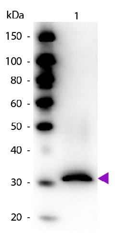 Western blot - Biotin Anti-RFP antibody (ab34771)Biotin Anti-RFP antibody (ab34771) at 1/1000 dilution
Secondary
Peroxidase streptavidin secondary antibody at 1/40000 dilution
-
 Immunohistochemistry (Frozen sections) - Biotin Anti-RFP antibody (ab34771) This image is courtesy of an anonymous Abreview
ab34771 staining mouse epidermis that had been damaged and injected with a mixture of cells inlcuding mouse embryonic stem cells previously transduced with a retrovirus that expresses RFP. The color is developed with DAB.
The sample was acetone fixed and incubated with ab34771 diluted 1/400 for 1 hour at 20°C. A Biotinylated goat anti-rabbit IgG (H+L) antibody was used as the secondary.


