Anti-YKL-40/CHI3L1 antibody (ab77528)
Key features and details
- Rabbit polyclonal to YKL-40/CHI3L1
- Suitable for: WB, IHC-P, ICC
- Reacts with: Human
- Isotype: IgG
Overview
-
Product name
Anti-YKL-40/CHI3L1 antibody
See all YKL-40/CHI3L1 primary antibodies -
Description
Rabbit polyclonal to YKL-40/CHI3L1 -
Host species
Rabbit -
Tested applications
Suitable for: WB, IHC-P, ICCmore details -
Species reactivity
Reacts with: Human
Predicted to work with: Mouse, Orangutan
-
Immunogen
Synthetic peptide corresponding to Human YKL-40/CHI3L1 aa 350 to the C-terminus (C terminal) conjugated to keyhole limpet haemocyanin.
(Peptide available asab88218) -
Positive control
- This antibody gave a positive signal in THP1 Whole Cell Lysate. This antibody gave a positive result in IHC in the following FFPE tissue: Human normal tonsil. This antibody gave a positive result when used in the following formaldehyde fixed cell lines: U-87 MG.
-
General notes
The Life Science industry has been in the grips of a reproducibility crisis for a number of years. Abcam is leading the way in addressing this with our range of recombinant monoclonal antibodies and knockout edited cell lines for gold-standard validation. Please check that this product meets your needs before purchasing.
If you have any questions, special requirements or concerns, please send us an inquiry and/or contact our Support team ahead of purchase. Recommended alternatives for this product can be found below, along with publications, customer reviews and Q&As
Properties
-
Form
Liquid -
Storage instructions
Shipped at 4°C. Store at +4°C short term (1-2 weeks). Upon delivery aliquot. Store at -20°C or -80°C. Avoid freeze / thaw cycle. -
Storage buffer
pH: 7.40
Preservative: 0.02% Sodium azide
Constituent: PBS
Batches of this product that have a concentration Concentration information loading...
Purity
Immunogen affinity purifiedClonality
PolyclonalIsotype
IgGResearch areas
Associated products
-
Compatible Secondaries
-
Isotype control
-
Recombinant Protein
Applications
The Abpromise guarantee
Our Abpromise guarantee covers the use of ab77528 in the following tested applications.
The application notes include recommended starting dilutions; optimal dilutions/concentrations should be determined by the end user.
Application Abreviews Notes WB (2) Use a concentration of 1 µg/ml. Detects a band of approximately 45 kDa (predicted molecular weight: 43 kDa).IHC-P Use a concentration of 1 µg/ml. Perform heat mediated antigen retrieval with citrate buffer pH 6 before commencing with IHC staining protocol.ICC Use a concentration of 5 µg/ml.Notes WB
Use a concentration of 1 µg/ml. Detects a band of approximately 45 kDa (predicted molecular weight: 43 kDa).IHC-P
Use a concentration of 1 µg/ml. Perform heat mediated antigen retrieval with citrate buffer pH 6 before commencing with IHC staining protocol.ICC
Use a concentration of 5 µg/ml.Target
-
Function
Carbohydrate-binding lectin with a preference for chitin. May play a role in defense against pathogens, or in tissue remodeling. May play an important role in the capacity of cells to respond to and cope with changes in their environment. -
Tissue specificity
Present in activated macrophages, articular chondrocytes, synovial cells as well as in liver. Undetectable in muscle tissues, lung, pancreas, mononuclear cells, or fibroblasts. -
Involvement in disease
A genetic variation in CHI3L1 is associated with susceptibility to asthma-related traits type 7 (ASRT7) [MIM:611960]. Asthma-related traits include clinical symptoms of asthma, such as coughing, wheezing and dyspnea, bronchial hyperresponsiveness (BHR) as assessed by methacholine challenge test, serum IgE levels, atopy, and atopic dermatitis. -
Sequence similarities
Belongs to the glycosyl hydrolase 18 family. -
Post-translational
modificationsGlycosylated. -
Cellular localization
Secreted > extracellular space. - Information by UniProt
-
Database links
- Entrez Gene: 1116 Human
- Entrez Gene: 12654 Mouse
- Omim: 601525 Human
- SwissProt: P36222 Human
- SwissProt: Q61362 Mouse
- Unigene: 382202 Human
- Unigene: 38274 Mouse
-
Alternative names
- 39 kDa synovial protein antibody
- ASRT7 antibody
- Cartilage glycoprotein 39 antibody
see all
Images
-
All lanes : Anti-YKL-40/CHI3L1 antibody (ab77528) at 1 µg/ml
Lane 1 : Wild-type THP-1 cell lysate
Lane 2 : CHI3L1 knockout THP-1 cell lysate
Lane 3 : U-87 MG cell lysate
Lane 4 : Jurkat cell lysate
Lysates/proteins at 20 µg per lane.
Performed under reducing conditions.
Predicted band size: 43 kDa
Observed band size: 33-42 kDa why is the actual band size different from the predicted?False colour image of Western blot: Anti-YKL-40/CHI3L1 antibody staining at 1 ug/ml, shown in green; Mouse anti-Alpha Tubulin [DM1A] (ab7291) loading control staining at 1/20000 dilution, shown in red. In Western blot, ab77528 was shown to bind specifically to YKL-40/CHI3L1. A band was observed at 33-42 kDa in wild-type THP-1 cell lysates with no signal observed at this size in CHI3L1 knockout cell line ab280038 (knockout cell lysate ab280097). To generate this image, wild-type and CHI3L1 knockout THP-1 cell lysates were analysed. First, samples were run on an SDS-PAGE gel then transferred onto a nitrocellulose membrane. Membranes were blocked in 3 % milk in TBS-0.1 % Tween® 20 (TBS-T) before incubation with primary antibodies overnight at 4 °C. Blots were washed four times in TBS-T, incubated with secondary antibodies for 1 h at room temperature, washed again four times then imaged. Secondary antibodies used were Goat anti-Rabbit IgG H&L (IRDye® 800CW) preabsorbed (ab216773) and Goat anti-Mouse IgG H&L (IRDye® 680RD) preabsorbed (ab216776) at 1/20000 dilution.
-
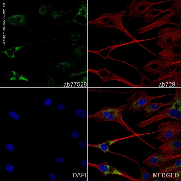
ab77528 staining Chitinase-3-like protein 1 precursor in U-87 MG cells. The cells were fixed with 100% methanol (5 min), permeabilized with 0.1% PBS-Triton X-100 for 5 minutes and then blocked with 1% BSA/10% normal goat serum/0.3M glycine in 0.1%PBS-Tween for 1h. The cells were then incubated overnight at 4°C with ab77528 at 5µg/ml and ab7291, Mouse monoclonal [DM1A] to alpha Tubulin - Loading Control. Cells were then incubated with ab150081, Goat polyclonal Secondary Antibody to Rabbit IgG - H&L (Alexa Fluor® 488), pre-adsorbed at 1/1000 dilution (shown in green) and ab150120, Goat polyclonal Secondary Antibody to Mouse IgG - H&L (Alexa Fluor® 594), pre-adsorbed at 1/1000 dilution (shown in pseudocolour red). Nuclear DNA was labelled with DAPI (shown in blue).
-
Anti-YKL-40/CHI3L1 antibody (ab77528) at 1 µg/ml + THP1 (Human acute monocytic leukemia cell line) Whole Cell Lysate at 10 µg
Secondary
Goat polyclonal to Rabbit IgG - H&L - Pre-Adsorbed (HRP) at 1/3000 dilution
Developed using the ECL technique.
Performed under reducing conditions.
Predicted band size: 43 kDa
Observed band size: 45 kDa why is the actual band size different from the predicted? -
 Immunohistochemistry (Formalin/PFA-fixed paraffin-embedded sections) - Anti-YKL-40/CHI3L1 antibody (ab77528)
IHC image of YKL-40/CHI3L1 staining in Human normal tonsil formalin fixed paraffin embedded tissue section, performed on a Leica Bond™ system using the standard protocol F. The section was pre-treated using heat mediated antigen retrieval with sodium citrate buffer (pH6, epitope retrieval solution 1) for 20 mins. The section was then incubated with ab77528, 1µg/ml, for 15 mins at room temperature and detected using an HRP conjugated compact polymer system. DAB was used as the chromogen. The section was then counterstained with haematoxylin and mounted with DPX.
For other IHC staining systems (automated and non-automated) customers should optimize variable parameters such as antigen retrieval conditions, primary antibody concentration and antibody incubation times.
Protocols
Datasheets and documents
-
SDS download
-
Datasheet download
References (18)
ab77528 has been referenced in 18 publications.
- Yu JE et al. Anti-Chi3L1 antibody suppresses lung tumor growth and metastasis through inhibition of M2 polarization. Mol Oncol 16:2214-2234 (2022). PubMed: 34861103
- Zhang S et al. SPI1-induced downregulation of FTO promotes GBM progression by regulating pri-miR-10a processing in an m6A-dependent manner. Mol Ther Nucleic Acids 27:699-717 (2022). PubMed: 35317283
- Zhao T et al. Chitinase-3 like-protein-1 promotes glioma progression via the NF-κB signaling pathway and tumor microenvironment reprogramming. Theranostics 12:6989-7008 (2022). PubMed: 36276655
- Christenson JL et al. Activity of Combined Androgen Receptor Antagonism and Cell Cycle Inhibition in Androgen Receptor Positive Triple Negative Breast Cancer. Mol Cancer Ther 20:1062-1071 (2021). PubMed: 33722849
- Chen Z et al. Cell surface GRP78 regulates BACE2 via lysosome-dependent manner to maintain mesenchymal phenotype of glioma stem cells. J Exp Clin Cancer Res 40:20 (2021). PubMed: 33413577
Images
-
ab77528 staining Chitinase-3-like protein 1 precursor in U-87 MG cells. The cells were fixed with 100% methanol (5 min), permeabilized with 0.1% PBS-Triton X-100 for 5 minutes and then blocked with 1% BSA/10% normal goat serum/0.3M glycine in 0.1%PBS-Tween for 1h. The cells were then incubated overnight at 4°C with ab77528 at 5µg/ml and ab7291, Mouse monoclonal [DM1A] to alpha Tubulin - Loading Control. Cells were then incubated with ab150081, Goat polyclonal Secondary Antibody to Rabbit IgG - H&L (Alexa Fluor® 488), pre-adsorbed at 1/1000 dilution (shown in green) and ab150120, Goat polyclonal Secondary Antibody to Mouse IgG - H&L (Alexa Fluor® 594), pre-adsorbed at 1/1000 dilution (shown in pseudocolour red). Nuclear DNA was labelled with DAPI (shown in blue).
-
Anti-YKL-40/CHI3L1 antibody (ab77528) at 1 µg/ml + THP1 (Human acute monocytic leukemia cell line) Whole Cell Lysate at 10 µg
Secondary
Goat polyclonal to Rabbit IgG - H&L - Pre-Adsorbed (HRP) at 1/3000 dilution
Developed using the ECL technique.
Performed under reducing conditions.
Predicted band size: 43 kDa
Observed band size: 45 kDa why is the actual band size different from the predicted?
-
 Immunohistochemistry (Formalin/PFA-fixed paraffin-embedded sections) - Anti-YKL-40/CHI3L1 antibody (ab77528)
IHC image of YKL-40/CHI3L1 staining in Human normal tonsil formalin fixed paraffin embedded tissue section, performed on a Leica Bond™ system using the standard protocol F. The section was pre-treated using heat mediated antigen retrieval with sodium citrate buffer (pH6, epitope retrieval solution 1) for 20 mins. The section was then incubated with ab77528, 1µg/ml, for 15 mins at room temperature and detected using an HRP conjugated compact polymer system. DAB was used as the chromogen. The section was then counterstained with haematoxylin and mounted with DPX.
For other IHC staining systems (automated and non-automated) customers should optimize variable parameters such as antigen retrieval conditions, primary antibody concentration and antibody incubation times.


