Anti-Vitamin D Binding protein antibody (ab65636)
")
Key features and details
- Rabbit polyclonal to Vitamin D Binding protein
- Suitable for: IHC-P, ICC/IF, WB
- Reacts with: Human
- Isotype: IgG
Overview
-
Product name
Anti-Vitamin D Binding protein antibody
See all Vitamin D Binding protein primary antibodies -
Description
Rabbit polyclonal to Vitamin D Binding protein -
Host species
Rabbit -
Tested applications
Suitable for: IHC-P, ICC/IF, WBmore details -
Species reactivity
Reacts with: Human
Predicted to work with: Pig
-
Immunogen
Synthetic peptide conjugated to KLH derived from within residues 450 to the C-terminus of Human Vitamin D Binding Protein.
Read Abcam's proprietary immunogen policy (Peptide available as ab71146.) -
Positive control
- WB: human liver and human ovary IHC-P: Human kidney FFPE tissue sections. ICC/IF: U937 cells
-
General notes
The Life Science industry has been in the grips of a reproducibility crisis for a number of years. Abcam is leading the way in addressing this with our range of recombinant monoclonal antibodies and knockout edited cell lines for gold-standard validation. Please check that this product meets your needs before purchasing.
If you have any questions, special requirements or concerns, please send us an inquiry and/or contact our Support team ahead of purchase. Recommended alternatives for this product can be found below, along with publications, customer reviews and Q&As
Properties
-
Form
Liquid -
Storage instructions
Shipped at 4°C. Store at +4°C short term (1-2 weeks). Upon delivery aliquot. Store at -20°C or -80°C. Avoid freeze / thaw cycle. -
Storage buffer
pH: 7.40
Preservative: 0.02% Sodium azide
Constituent: PBS
Batches of this product that have a concentration Concentration information loading...
Purity
Immunogen affinity purifiedClonality
PolyclonalIsotype
IgGResearch areas
Associated products
-
Compatible Secondaries
-
Immunizing Peptide (Blocking)
-
Isotype control
-
Recombinant Protein
Applications
The Abpromise guarantee
Our Abpromise guarantee covers the use of ab65636 in the following tested applications.
The application notes include recommended starting dilutions; optimal dilutions/concentrations should be determined by the end user.
Application Abreviews Notes IHC-P Use a concentration of 1 µg/ml.ICC/IF Use a concentration of 5 µg/ml.WB Use a concentration of 1 µg/ml. Detects a band of approximately 53 kDa (predicted molecular weight: 53 kDa).Notes IHC-P
Use a concentration of 1 µg/ml.ICC/IF
Use a concentration of 5 µg/ml.WB
Use a concentration of 1 µg/ml. Detects a band of approximately 53 kDa (predicted molecular weight: 53 kDa).Target
-
Function
Multifunctional protein found in plasma, ascitic fluid, cerebrospinal fluid, and urine and on the surface of many cell types. In plasma, it carries the vitamin D sterols and prevents polymerization of actin by binding its monomers. DBP associates with membrane-bound immunoglobulin on the surface of B-lymphocytes and with IgG Fc receptor on the membranes of T-lymphocytes. -
Sequence similarities
Belongs to the ALB/AFP/VDB family.
Contains 3 albumin domains. -
Cellular localization
Secreted. - Information by UniProt
-
Database links
- Entrez Gene: 2638 Human
- Omim: 139200 Human
- SwissProt: P02774 Human
- Unigene: 418497 Human
-
Alternative names
- DBP antibody
- DBP/GC antibody
- GC antibody
see all
Images
-
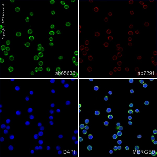 Immunocytochemistry/ Immunofluorescence - Anti-Vitamin D Binding protein antibody (ab65636)
ab65636 staining Vitamin D binding protein in U937 cells. The cells were fixed with 100% methanol (5 min), permeabilized with 0.1% PBS-Tween for 5 minutes and then blocked with 1% BSA/10% normal goat serum/0.3M glycine in 0.1% PBS-Tween for 1h. The cells were then incubated overnight at 4°C with ab65636 at 5µg/ml and ab7291, Mouse monoclonal [DM1A] to alpha Tubulin - Loading Control. Cells were then incubated with ab150081, Goat polyclonal Secondary Antibody to Rabbit IgG - H&L (Alexa Fluor® 488), pre-adsorbed at 1/1000 dilution (shown in green) and ab150080, Goat polyclonal Secondary Antibody to Rabbit IgG - H&L (Alexa Fluor® 594) at 1/1000 dilution (shown in pseudocolour red). Nuclear DNA was labelled with DAPI (shown in blue).
Image was acquired with a confocal microscope (Leica-Microsystems TCS SP8) and a single confocal section is shown.
-
Western blot - Anti-Vitamin D Binding protein antibody (ab65636)All lanes : Anti-Vitamin D Binding protein antibody (ab65636) at 1 µg/ml
Lane 1 : Human liver tissue lysate - total protein (ab29889)
Lane 2 : Human ovary tissue lysate - total protein (ab30222)
Lysates/proteins at 10 µg per lane.
Secondary
All lanes : Goat polyclonal to Rabbit IgG - H&L - Pre-Adsorbed (HRP) at 1/3000 dilution
Performed under reducing conditions.
Predicted band size: 53 kDa
Observed band size: 53 kDa
Exposure time: 1 minute -
 Immunohistochemistry (Formalin/PFA-fixed paraffin-embedded sections) - Anti-Vitamin D Binding protein antibody (ab65636)
IHC image of Vitamin D Binding Protein staining in human kidney FFPE section, performed on a BondTM system using the standard protocol F. The section was pre-treated using heat mediated antigen retrieval with sodium citrate buffer (pH6, epitope retrieval solution 1) for 20 mins. The section was then incubated with ab65636, 1µg/ml, for 15 mins at room temperature and detected using an HRP conjugated compact polymer system. DAB was used as the chromogen. The section was then counterstained with haematoxylin and mounted with DPX
-
 Western blot - Anti-Vitamin D Binding protein antibody (ab65636)
Protocols
Datasheets and documents
-
SDS download
-
Datasheet download
References (5)
ab65636 has been referenced in 5 publications.
- Lam CJ et al. Low-Level Insulin Content Within Abundant Non-ß Islet Endocrine Cells in Long-standing Type 1 Diabetes. Diabetes 68:598-608 (2019). PubMed: 30552110
- Ackermann AM et al. Integration of ATAC-seq and RNA-seq identifies human alpha cell and beta cell signature genes. Mol Metab 5:233-44 (2016). Human . PubMed: 26977395
- Taniuchi K et al. RUVBL1 directly binds actin filaments and induces formation of cell protrusions to promote pancreatic cancer cell invasion. Int J Oncol 44:1945-54 (2014). PubMed: 24728183
- Tao W et al. Hormonal induction and roles of Disabled-2 in lactation and involution. PLoS One 9:e110737 (2014). WB ; Mouse . PubMed: 25360623
- Hernández-Martín A et al. Synthesis of vitamin D3 analogues with A-ring modifications to directly measure vitamin D levels in biological samples. Bioorg Med Chem 21:7779-89 (2013). PubMed: 24216092
Images
-
Western blot - Anti-Vitamin D Binding protein antibody (ab65636)All lanes : Anti-Vitamin D Binding protein antibody (ab65636) at 1 µg/ml
Lane 1 : Human liver tissue lysate - total protein (ab29889)
Lane 2 : Human ovary tissue lysate - total protein (ab30222)
Lysates/proteins at 10 µg per lane.
Secondary
All lanes : Goat polyclonal to Rabbit IgG - H&L - Pre-Adsorbed (HRP) at 1/3000 dilution
Performed under reducing conditions.
Predicted band size: 53 kDa
Observed band size: 53 kDa
Exposure time: 1 minute
-
Immunohistochemistry (Formalin/PFA-fixed paraffin-embedded sections) - Anti-Vitamin D Binding protein antibody (ab65636)
IHC image of Vitamin D Binding Protein staining in human kidney FFPE section, performed on a BondTM system using the standard protocol F. The section was pre-treated using heat mediated antigen retrieval with sodium citrate buffer (pH6, epitope retrieval solution 1) for 20 mins. The section was then incubated with ab65636, 1µg/ml, for 15 mins at room temperature and detected using an HRP conjugated compact polymer system. DAB was used as the chromogen. The section was then counterstained with haematoxylin and mounted with DPX
-
Western blot - Anti-Vitamin D Binding protein antibody (ab65636)
-
 Immunocytochemistry/ Immunofluorescence - Anti-Vitamin D Binding protein antibody (ab65636)ICC/IF image of ab65636 stained HeLa cells. The cells were 4% PFA fixed (10 min) and then incubated in 1%BSA / 10% normal goat serum / 0.3M glycine in 0.1% PBS-Tween for 1h to permeabilise the cells and block non-specific protein-protein interactions. The cells were then incubated with the antibody (ab65636, 5µg/ml) overnight at +4°C. The secondary antibody (green) was Alexa Fluor® 488 goat anti-rabbit IgG (H+L) used at a 1/1000 dilution for 1h. Alexa Fluor® 594 WGA was used to label plasma membranes (red) at a 1/200 dilution for 1h. DAPI was used to stain the cell nuclei (blue) at a concentration of 1.43µM.


