Anti-Survivin antibody (ab469)
Key features and details
- Rabbit polyclonal to Survivin
- Suitable for: ICC/IF, WB, IHC-P
- Reacts with: Human
- Isotype: IgG
Overview
-
Product name
Anti-Survivin antibody
See all Survivin primary antibodies -
Description
Rabbit polyclonal to Survivin -
Host species
Rabbit -
Specificity
Specific for survivin. -
Tested applications
Suitable for: ICC/IF, WB, IHC-Pmore details -
Species reactivity
Reacts with: Human -
Immunogen
Recombinant full length protein corresponding to Human Survivin .
Database link: O15392
Properties
-
Form
Liquid -
Storage instructions
Shipped at 4°C. Store at +4°C short term (1-2 weeks). Upon delivery aliquot. Store at -20°C long term. Avoid freeze / thaw cycle. -
Storage buffer
pH: 7.40
Preservative: 0.05% Sodium azide
Constituents: 0.876% Sodium chloride, 99% Tris glycine -
 Concentration information loading...
 -
Purity
Immunogen affinity purified -
Clonality
Polyclonal -
Isotype
IgG -
Research areas
Images
-
All lanes : Anti-Survivin antibody (ab469) at 1 µg/ml
Lane 1 : HeLa Nuclear
Lane 2 : HeLa whole cell lysate
Lane 3 : A431 cell lysate
Lane 4 : Jurkat cell lysate
Lane 5 : HEK293 cell lysate
Lysates/proteins at 20 µg per lane.
Secondary
All lanes : Alexa Fluor anti-rabbit at 1/5000 dilution
Performed under reducing conditions.
Predicted band size: 16 kDa
Observed band size: 18 kDa why is the actual band size different from the predicted?
Additional bands at: 37 kDa, 50 kDa. We are unsure as to the identity of these extra bands.
-
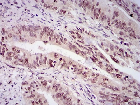
 Immunohistochemistry (Formalin/PFA-fixed paraffin-embedded sections) - Anti-Survivin antibody (ab469)
Paraffin-embedded human rectal cancer tissue stained for Survivin using ab469 at 0.5 µg/ml in immunohistochemical analysis, using DAB with hematoxylin counterstain.
-
HeLa (human epithelial cell line from cervix adenocarcinoma) cells stained for Survivin (green) using ab469 at 1/10 dilution in ICC/IF. An Alexa Fluor 488-conjugated Goat to rabbit IgG was used as secondary antibody (green). Actin filaments were labeled with Alexa Fluor 568 phalloidin (red). DAPI was used to stain the cell nuclei (blue).
-
 Immunohistochemistry (Formalin/PFA-fixed paraffin-embedded sections) - Anti-Survivin antibody (ab469) This image is courtesy of an Abreview submitted by Dr Ben Davidson
ab469 staining Survivin from Human Ovarian carcionoma tumour tissue sections by Immunohistochemistry (Formalin-fixed paraffin-embedded sections). Heat mediated antigen retrieval was performed (Citrate buffer pH=6, microwave oven) and the tissue was then formaldehyde fixed and blocked (Hydrogen peroxide 0.03%). An HRP conjugated goat anti-rabbit was used as the secondary antibody.
-
 Immunocytochemistry/ Immunofluorescence - Anti-Survivin antibody (ab469) This image is courtesy of William Moore
HeLa cells (ab150035) in prometaphase, metaphase and anaphase stained with anti-Survivin (green), anti-tubulin (red) and DAPI (blue). These images were kindly supplied as part of the review submitted by William Moore, University of Dundee, UK.
-
ab469 at a 1/400 dilution staining HeLa cells by Immunocytochemistry. The antibody was incubated with the cells for 1 hour and then was detected using a Texas Red conjugated Goat anti-rabbit antibody.
This image is courtesy of an Abreview by Sandrine Ruchaud submitted on 30 March 2006.
-
Anti-Survivin antibody (ab469) at 1 µg/ml + HeLa (human epithelial cell line from cervix adenocarcinoma) whole cell lysate at 30 µg
Developed using the ECL technique.
Predicted band size: 16 kDa
Exposure time: 1 minute
-
 Immunohistochemistry (Formalin/PFA-fixed paraffin-embedded sections) - Anti-Survivin antibody (ab469) This image is courtesy of an Abreview submitted by Mr. Rudolf Jung.Paraformaldehyde-fixed, paraffin-embedded human colon carcinoma tissue stained for Survivin using ab469 at 1/500 dilution in immunohistochemical analysis.


