Anti-Sumo 1 antibody (ab49767)
Key features and details
- Rabbit polyclonal to Sumo 1
- Suitable for: WB, ICC/IF
- Knockout validated
- Reacts with: Human
- Isotype: IgG
Overview
-
Product name
Anti-Sumo 1 antibody
See all Sumo 1 primary antibodies -
Description
Rabbit polyclonal to Sumo 1 -
Host species
Rabbit -
Tested Applications & Species
See all applications and species dataApplication Species ICC/IF HumanWB Human -
Immunogen
Synthetic peptide corresponding to Human Sumo 1 aa 1-100 conjugated to keyhole limpet haemocyanin.
(Peptide available asab49766) -
Positive control
- Recombinant Human Sumo 1 protein (ab84193) can be used as a positive control in WB. This antibody gave a positive signal in the following whole cell lyates: A431; Jurkat (data not shown)
Properties
-
Form
Liquid -
Storage instructions
Shipped at 4°C. Store at +4°C short term (1-2 weeks). Upon delivery aliquot. Store at -20°C or -80°C. Avoid freeze / thaw cycle. -
Storage buffer
pH: 7.40
Preservative: 0.02% Sodium azide
Constituent: PBS
Batches of this product that have a concentration Concentration information loading...
Purity
Immunogen affinity purifiedClonality
PolyclonalIsotype
IgGResearch areas
Associated products
-
Compatible Secondaries
-
Isotype control
-
Recombinant Protein
Applications
The Abpromise guarantee
Our Abpromise guarantee covers the use of ab49767 in the following tested applications.
The application notes include recommended starting dilutions; optimal dilutions/concentrations should be determined by the end user.
GuaranteedTested applications are guaranteed to work and covered by our Abpromise guarantee.
PredictedPredicted to work for this combination of applications and species but not guaranteed.
IncompatibleDoes not work for this combination of applications and species.
Application Species ICC/IF HumanWB HumanAll applications MouseRatCowPigOrangutanApplication Abreviews Notes WB Use a concentration of 1 µg/ml. Detects a band of approximately 16, 80 kDa (predicted molecular weight: 12 kDa).ICC/IF Use a concentration of 5 µg/ml.Notes WB
Use a concentration of 1 µg/ml. Detects a band of approximately 16, 80 kDa (predicted molecular weight: 12 kDa).ICC/IF
Use a concentration of 5 µg/ml.Target
-
Function
Ubiquitin-like protein that can be covalently attached to proteins as a monomer or a lysine-linked polymer. Covalent attachment via an isopeptide bond to its substrates requires prior activation by the E1 complex SAE1-SAE2 and linkage to the E2 enzyme UBE2I, and can be promoted by E3 ligases such as PIAS1-4, RANBP2 or CBX4. This post-translational modification on lysine residues of proteins plays a crucial role in a number of cellular processes such as nuclear transport, DNA replication and repair, mitosis and signal transduction. Involved for instance in targeting RANGAP1 to the nuclear pore complex protein RANBP2. Polymeric SUMO1 chains are also susceptible to polyubiquitination which functions as a signal for proteasomal degradation of modified proteins. May also regulate a network of genes involved in palate development. -
Involvement in disease
Defects in SUMO1 are the cause of non-syndromic orofacial cleft type 10 (OFC10) [MIM:613705]; also called non-syndromic cleft lip with or without cleft palate 10. OFC10 is a birth defect consisting of cleft lips with or without cleft palate. Cleft lips are associated with cleft palate in two-third of cases. A cleft lip can occur on one or both sides and range in severity from a simple notch in the upper lip to a complete opening in the lip extending into the floor of the nostril and involving the upper gum. Note=A chromosomal aberation involving SUMO1 is the cause of OFC10. Translocation t(2;8)(q33.1;q24.3). The breakpoint occurred in the SUMO1 gene and resulted in haploinsufficiency confirmed by protein assays. -
Sequence similarities
Belongs to the ubiquitin family. SUMO subfamily.
Contains 1 ubiquitin-like domain. -
Post-translational
modificationsCleavage of precursor form by SENP1 or SENP2 is necessary for function.
Polymeric SUMO1 chains undergo polyubiquitination by RNF4. -
Cellular localization
Nucleus membrane. Nucleus speckle. Cytoplasm. Recruited by BCL11A into the nuclear body. - Information by UniProt
-
Database links
- Entrez Gene: 614967 Cow
- Entrez Gene: 7341 Human
- Entrez Gene: 22218 Mouse
- Entrez Gene: 100173521 Orangutan
- Entrez Gene: 100127139 Pig
- Entrez Gene: 301442 Rat
- Omim: 601912 Human
- SwissProt: Q5E9D1 Cow
see all -
Alternative names
- DAP1 antibody
- GAP modifying protein 1 antibody
- GAP-modifying protein 1 antibody
see all
Images
-
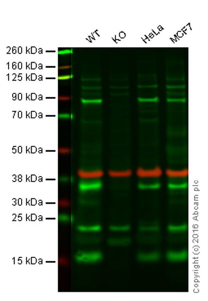
Lane 1: Wild-type HAP1 cell lysate (20 µg)
Lane 2: Sumo 1 knockout HAP1 cell lysate (20 µg)
Lane 3: HeLa cell lysate (20 µg)
Lane 4: MCF-7 cell lysate (20 µg)
Lanes 1 - 4: Merged signal (red and green). Green - ab49767 observed at 16 kDa. Red - loading control, ab8245, observed at 37kDa.
ab49767 was shown to recognize Sumo 1 when Sumo 1 knockout samples were used, along with additional cross-reactive bands. Wild-type and Sumo 1 knockout samples were subjected to SDS-PAGE. ab49767 at a concentration of 1 μg/ml and ab8245 (loading control to GAPDH) at a dilution of 1/10000 were incubated overnight at 4°C. Blots were developed with Goat anti-Rabbit IgG H&L (IRDye® 800CW) preadsorbed ab216773 and Goat anti-Mouse IgG H&L (IRDye® 680RD) preadsorbed ab216776 secondary antibodies at 1/10000 dilution for 1 hour at room temperature before imaging. -
Anti-Sumo 1 antibody (ab49767) at 1 µg/ml + A431 (Human epithelial carcinoma cell line) Whole Cell Lysate at 10 µg
Secondary
IRDye 680 Conjugated Goat Anti-Rabbit IgG (H+L) at 1/10000 dilution
Performed under reducing conditions.
Predicted band size: 12 kDa
Observed band size: 16,80 kDa why is the actual band size different from the predicted?
Sumo 1 is known to form a complex with RanGAP, resulting in the band seen at 80 kDa -
ICC/IF image of ab49767 stained HeLa cells. The cells were 4% PFA fixed (10 min) and then incubated in 1%BSA / 10% normal goat serum / 0.3M glycine in 0.1% PBS-Tween for 1h to permeabilise the cells and block non-specific protein-protein interactions. The cells were then incubated with the antibody (ab49767, 5µg/ml) overnight at +4°C. The secondary antibody (green) was Alexa Fluor® 488 goat anti-rabbit IgG (H+L) used at a 1/1000 dilution for 1h. Alexa Fluor® 594 WGA was used to label plasma membranes (red) at a 1/200 dilution for 1h. DAPI was used to stain the cell nuclei (blue) at a concentration of 1.43µM. This antibody also gave a positive result in 4% PFA fixed (10 min) Hek293, HepG2 and MCF7 cells at 5µg/ml, and in 100% methanol fixed (5 min) HeLa, Hek293, HepG2 and MCF7 cells at 5µg/ml.
Protocols
Datasheets and documents
References (1)
ab49767 has been referenced in 1 publication.
- Chowdhury D et al. p38 MAPK pathway-dependent SUMOylation of Elk-1 and phosphorylation of PIAS2 correlate with the downregulation of Elk-1 activity in heat-stressed HeLa cells. Cell Stress Chaperones 24:393-407 (2019). PubMed: 30783905
Images
-
Lane 1: Wild-type HAP1 cell lysate (20 µg)
Lane 2: Sumo 1 knockout HAP1 cell lysate (20 µg)
Lane 3: HeLa cell lysate (20 µg)
Lane 4: MCF-7 cell lysate (20 µg)
Lanes 1 - 4: Merged signal (red and green). Green - ab49767 observed at 16 kDa. Red - loading control, ab8245, observed at 37kDa.
ab49767 was shown to recognize Sumo 1 when Sumo 1 knockout samples were used, along with additional cross-reactive bands. Wild-type and Sumo 1 knockout samples were subjected to SDS-PAGE. ab49767 at a concentration of 1 μg/ml and ab8245 (loading control to GAPDH) at a dilution of 1/10000 were incubated overnight at 4°C. Blots were developed with Goat anti-Rabbit IgG H&L (IRDye® 800CW) preadsorbed ab216773 and Goat anti-Mouse IgG H&L (IRDye® 680RD) preadsorbed ab216776 secondary antibodies at 1/10000 dilution for 1 hour at room temperature before imaging. -
Anti-Sumo 1 antibody (ab49767) at 1 µg/ml + A431 (Human epithelial carcinoma cell line) Whole Cell Lysate at 10 µg
Secondary
IRDye 680 Conjugated Goat Anti-Rabbit IgG (H+L) at 1/10000 dilution
Performed under reducing conditions.
Predicted band size: 12 kDa
Observed band size: 16,80 kDa why is the actual band size different from the predicted?
Sumo 1 is known to form a complex with RanGAP, resulting in the band seen at 80 kDa -
ICC/IF image of ab49767 stained HeLa cells. The cells were 4% PFA fixed (10 min) and then incubated in 1%BSA / 10% normal goat serum / 0.3M glycine in 0.1% PBS-Tween for 1h to permeabilise the cells and block non-specific protein-protein interactions. The cells were then incubated with the antibody (ab49767, 5µg/ml) overnight at +4°C. The secondary antibody (green) was Alexa Fluor® 488 goat anti-rabbit IgG (H+L) used at a 1/1000 dilution for 1h. Alexa Fluor® 594 WGA was used to label plasma membranes (red) at a 1/200 dilution for 1h. DAPI was used to stain the cell nuclei (blue) at a concentration of 1.43µM. This antibody also gave a positive result in 4% PFA fixed (10 min) Hek293, HepG2 and MCF7 cells at 5µg/ml, and in 100% methanol fixed (5 min) HeLa, Hek293, HepG2 and MCF7 cells at 5µg/ml.


