Anti-OPA1 antibody (ab42364)
Key features and details
- Rabbit polyclonal to OPA1
- Suitable for: WB, IP
- Reacts with: Mouse, Rat, Human
- Isotype: IgG
Overview
-
Product name
Anti-OPA1 antibody
See all OPA1 primary antibodies -
Description
Rabbit polyclonal to OPA1 -
Host species
Rabbit -
Tested applications
Suitable for: WB, IPmore details -
Species reactivity
Reacts with: Mouse, Rat, Human
Predicted to work with: Chicken, Orangutan
-
Immunogen
Synthetic peptide within Rat OPA1 aa 900 to the C-terminus (C terminal) conjugated to keyhole limpet haemocyanin. The exact sequence is proprietary.
(Peptide available asab42363)
Properties
-
Form
Liquid -
Storage instructions
Shipped at 4°C. Store at +4°C short term (1-2 weeks). Upon delivery aliquot. Store at -20°C or -80°C. Avoid freeze / thaw cycle. -
Storage buffer
pH: 7.40
Preservative: 0.02% Sodium azide
Constituent: PBS
Batches of this product that have a concentration Concentration information loading...
Purity
Immunogen affinity purifiedClonality
PolyclonalIsotype
IgGResearch areas
Associated products
-
Compatible Secondaries
-
Isotype control
-
Recombinant Protein
Applications
Our Abpromise guarantee covers the use of ab42364 in the following tested applications.
The application notes include recommended starting dilutions; optimal dilutions/concentrations should be determined by the end user.
Application Abreviews Notes WB Use a concentration of 1 µg/ml. Detects a band of approximately 92,86 kDa (predicted molecular weight: 111 kDa). Abcam recommends using milk as the blocking agent.
IP Use at an assay dependent concentration. Target
-
Function
Dynamin-related GTPase required for mitochondrial fusion and regulation of apoptosis. May form a diffusion barrier for proteins stored in mitochondrial cristae. Proteolytic processing in response to intrinsic apoptotic signals may lead to disassembly of OPA1 oligomers and release of the caspase activator cytochrome C (CYCS) into the mitochondrial intermembrane space. -
Tissue specificity
Highly expressed in retina. Also expressed in brain, testis, heart and skeletal muscle. Isoform 1 expressed in retina, skeletal muscle, heart, lung, ovary, colon, thyroid gland, leukocytes and fetal brain. Isoform 2 expressed in colon, liver, kidney, thyroid gland and leukocytes. Low levels of all isoforms expressed in a variety of tissues. -
Involvement in disease
Defects in OPA1 are a cause of optic atrophy type 1 (OPA1) [MIM:165500]. OPA1 is a dominantly inherited optic neuropathy occurring in 1 in 50,000 individuals that features progressive loss in visual acuity leading, in many cases, to legal blindness.
Defects in OPA1 are the cause of optic atrophy 1 with deafness (OPA1D) [MIM:125250]. Some individuals with mutations in OPA1 manifest also ophthalmoplegia and myopathy. -
Sequence similarities
Belongs to the dynamin family. -
Post-translational
modificationsPARL-dependent proteolytic processing releases an antiapoptotic soluble form not required for mitochondrial fusion. -
Cellular localization
Mitochondrion inner membrane. Mitochondrion intermembrane space. - Information by UniProt
-
Database links
- Entrez Gene: 424900 Chicken
- Entrez Gene: 4976 Human
- Entrez Gene: 74143 Mouse
- Entrez Gene: 171116 Rat
- Omim: 605290 Human
- SwissProt: O60313 Human
- SwissProt: P58281 Mouse
- SwissProt: Q2TA68 Rat
see all -
Alternative names
- Dynamin like 120 kDa protein antibody
- Dynamin like 120 kDa protein, mitochondrial antibody
- Dynamin-like 120 kDa protein, form S1 antibody
see all
Images
-
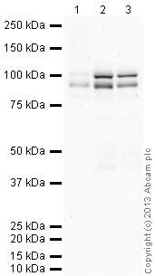
All lanes : Anti-OPA1 antibody (ab42364) at 1 µg/ml
Lane 1 : Human brain tissue lysate - total protein (ab29466)
Lane 2 : Brain (Mouse) Tissue Lysate
Lane 3 : Brain (Rat) Tissue Lysate
Lysates/proteins at 10 µg per lane.
Secondary
All lanes : Goat Anti-Rabbit IgG H&L (HRP) (ab97051) at 1/10000 dilution
Developed using the ECL technique.
Performed under reducing conditions.
Predicted band size: 111 kDa
Observed band size: 92 kDa why is the actual band size different from the predicted?
Additional bands at: 100 kDa. We are unsure as to the identity of these extra bands.
Exposure time: 2 minutesAbcam recommends using milk as the blocking agent. Abcam welcomes customer feedback and would appreciate any comments regarding this product and the data presented above.
-
Anti-OPA1 antibody (ab42364) at 1 µg/ml + Brain (Rat) Tissue Lysate - normal tissue at 10 µg
Secondary
Goat polyclonal to Rabbit IgG - H&L - Pre-Adsorbed (HRP) at 1/3000 dilution
Performed under reducing conditions.
Predicted band size: 111 kDa
Observed band size: 92 kDa why is the actual band size different from the predicted?
Additional bands at: 86 kDa (possible isoform)
Although the predicted band size is ~111 kDa based on Swiss-prot data, bands of 92 kDa and 86 kDa has been previously observed. Investigative Ophthalmology & Visual Science, November 2005, Vol. 46, No. 11 -
OPA1 was immunoprecipitated using 0.5mg Rat Brain whole tissue lysate, 5µg of Rabbit polyclonal to OPA1 and 50µl of protein G magnetic beads (+). No antibody was added to the control (-).
The antibody was incubated under agitation with Protein G beads for 10min, rat brain whole tissue lysate diluted in RIPA buffer was added to each sample and incubated for a further 10min under agitation.
Proteins were eluted by addition of 40µl SDS loading buffer and incubated for 10min at 70oC; 10µl of each sample was separated on a SDS PAGE gel, transferred to a nitrocellulose membrane, blocked with 5% BSA and probed with ab42364.
Secondary: Mouse monoclonal [SB62a] Secondary Antibody to Rabbit IgG light chain (HRP) (ab99697).
Band: 92kDa: OPA1; non specific - 100kDa: We are unsure as to the identity of this extra band.
Protocols
References (86)
ab42364 has been referenced in 86 publications.
- Gokita K et al. Therapeutic Potential of LNP-Mediated Delivery of miR-634 for Cancer Therapy. Mol Ther Nucleic Acids 19:330-338 (2020). PubMed: 31877409
- Kang L et al. The mitochondria-targeted anti-oxidant MitoQ protects against intervertebral disc degeneration by ameliorating mitochondrial dysfunction and redox imbalance. Cell Prolif 53:e12779 (2020). PubMed: 32020711
- Chen D et al. Systematic analysis of a mitochondrial disease-causing ND6 mutation in mitochondrial deficiency. Mol Genet Genomic Med 8:e1199 (2020). PubMed: 32162843
- Chen WR et al. Melatonin Attenuates Calcium Deposition from Vascular Smooth Muscle Cells by Activating Mitochondrial Fusion and Mitophagy via an AMPK/OPA1 Signaling Pathway. Oxid Med Cell Longev 2020:5298483 (2020). PubMed: 32377301
- Yuan Q et al. Role of pyruvate kinase M2-mediated metabolic reprogramming during podocyte differentiation. Cell Death Dis 11:355 (2020). PubMed: 32393782
Images
-
All lanes : Anti-OPA1 antibody (ab42364) at 1 µg/ml
Lane 1 : Human brain tissue lysate - total protein (ab29466)
Lane 2 : Brain (Mouse) Tissue Lysate
Lane 3 : Brain (Rat) Tissue Lysate
Lysates/proteins at 10 µg per lane.
Secondary
All lanes : Goat Anti-Rabbit IgG H&L (HRP) (ab97051) at 1/10000 dilution
Developed using the ECL technique.
Performed under reducing conditions.
Predicted band size: 111 kDa
Observed band size: 92 kDa why is the actual band size different from the predicted?
Additional bands at: 100 kDa. We are unsure as to the identity of these extra bands.
Exposure time: 2 minutesAbcam recommends using milk as the blocking agent. Abcam welcomes customer feedback and would appreciate any comments regarding this product and the data presented above.
-
Anti-OPA1 antibody (ab42364) at 1 µg/ml + Brain (Rat) Tissue Lysate - normal tissue at 10 µg
Secondary
Goat polyclonal to Rabbit IgG - H&L - Pre-Adsorbed (HRP) at 1/3000 dilution
Performed under reducing conditions.
Predicted band size: 111 kDa
Observed band size: 92 kDa why is the actual band size different from the predicted?
Additional bands at: 86 kDa (possible isoform)
Although the predicted band size is ~111 kDa based on Swiss-prot data, bands of 92 kDa and 86 kDa has been previously observed. Investigative Ophthalmology & Visual Science, November 2005, Vol. 46, No. 11 -
OPA1 was immunoprecipitated using 0.5mg Rat Brain whole tissue lysate, 5µg of Rabbit polyclonal to OPA1 and 50µl of protein G magnetic beads (+). No antibody was added to the control (-).
The antibody was incubated under agitation with Protein G beads for 10min, rat brain whole tissue lysate diluted in RIPA buffer was added to each sample and incubated for a further 10min under agitation.
Proteins were eluted by addition of 40µl SDS loading buffer and incubated for 10min at 70oC; 10µl of each sample was separated on a SDS PAGE gel, transferred to a nitrocellulose membrane, blocked with 5% BSA and probed with ab42364.
Secondary: Mouse monoclonal [SB62a] Secondary Antibody to Rabbit IgG light chain (HRP) (ab99697).
Band: 92kDa: OPA1; non specific - 100kDa: We are unsure as to the identity of this extra band.


