Anti-GRP94 antibody (ab3674)
")
Key features and details
- Rabbit polyclonal to GRP94
- Suitable for: WB, IHC-P, ICC/IF
- Reacts with: Mouse, Human
- Isotype: IgG
Properties
-
Form
Liquid -
Storage instructions
Shipped at 4°C. Store at +4°C short term (1-2 weeks). Upon delivery aliquot. Store at -20°C or -80°C. Avoid freeze / thaw cycle. -
Storage buffer
Preservative: 0.01% Sodium azide -
 Concentration information loading...
 -
Purity
Whole antiserum -
Clonality
Polyclonal -
Isotype
IgG -
Research areas
Images
-
Immunocytochemistry/ Immunofluorescence - Anti-GRP94 antibody (ab3674)Immunostaining of mouse embryonic fibroblasts with ab3674. (See Nakamura et al. 2001).
-
 Western blot - Anti-GRP94 antibody (ab3674)Anti-GRP94 antibody (ab3674) at 1/2000 dilution + Cell lysates prepared from mouse fibroblasts
Western blot of mouse fibroblasts using ab3764 at 1/2000. -
 Immunocytochemistry/ Immunofluorescence - Anti-GRP94 antibody (ab3674) This image is courtesy of an anonymous Abreviewab3674 at a 1/100 dilution staining human renal cell carcinoma cells by ICC/IF. The cells were permeabilized with 0.2% triton X100 and then blocked with 1%BSA, 4% goat serum prior to incubation with the primary 1antibody for 30 min at 37 °C. Bound antibody was detected using an Alexa fluor ® 488 Goat anti-Rabbit IgG (H+L).
 -
 Immunohistochemistry (Formalin/PFA-fixed paraffin-embedded sections) - Anti-GRP94 antibody (ab3674)
Ab3674 (1/1000) staining GPR94 in human stomach using an automated system (DAKO Autostainer Plus). Using this protocol there is strong cytoplasmic staining of peptic and parietal cells of the gastric gland.
Sections were rehydrated and antigen retrieved with the Dako 3 in 1 AR buffer EDTA pH 9.0 in a DAKO PT link. Slides were peroxidase blocked in 3% H2O2 in methanol for 10 mins. They were then blocked with Dako Protein block for 10 minutes (containing casein 0.25% in PBS) then incubated with primary antibody for 20 min and detected with Dako envision flex amplification kit for 30 minutes. Colorimetric detection was completed with Diaminobenzidine for 5 minutes. Slides were counterstained with Haematoxylin and coverslipped under DePeX. Please note that, for manual staining, optimization of primary antibody concentration and incubation time is recommended. Signal amplification may be required. -
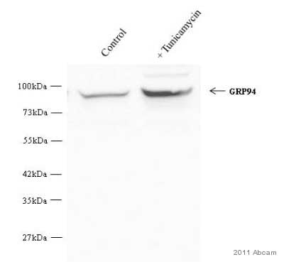 Western blot - Anti-GRP94 antibody (ab3674) Image courtesy of an anonymous Abreview.All lanes : Anti-GRP94 antibody (ab3674) at 1/2000 dilution
Lane 1 : Whole cell lysate prepared from murine 3T3 cells
Lane 2 : Whole cell lysate prepared from murine 3T3 cells, treated with 2.5 ug/ml Tunicamycin 10 hours
Lysates/proteins at 50 µg per lane.
Secondary
All lanes : HRP conjugated goat anti-rabbit IgG at 1/2000 dilution
Developed using the ECL technique.
Observed band size: 94 kDa why is the actual band size different from the predicted?
Exposure time: 30 seconds


