Anti-GRP78 BiP antibody (ab32618)
")
Key features and details
- Rabbit polyclonal to GRP78 BiP
- Suitable for: WB, IHC-P, ICC/IF
- Reacts with: Mouse, Human, Chinese hamster
- Isotype: IgG
Overview
-
Product name
Anti-GRP78 BiP antibody
See all GRP78 BiP primary antibodies -
Description
Rabbit polyclonal to GRP78 BiP -
Host species
Rabbit -
Tested applications
Suitable for: WB, IHC-P, ICC/IFmore details -
Species reactivity
Reacts with: Mouse, Human, Chinese hamster
Predicted to work with: Rat, Chicken, Hamster, Xenopus laevis
-
Immunogen
Synthetic peptide within Human GRP78 BiP aa 1-100. The exact sequence is proprietary.
Database link: P11021 -
Positive control
- HeLa cells, breast carcinoma.
-
General notes
This product is FOR RESEARCH USE ONLY. For commercial use, please contact partnerships@abcam.com.
Properties
-
Form
Liquid -
Storage instructions
Shipped at 4°C. Upon delivery aliquot and store at -20°C. Avoid freeze / thaw cycles. -
Storage buffer
pH: 7.60
Preservative: 0.1% Sodium azide
Constituents: PBS, 1% BSA -
 Concentration information loading...
 -
Purity
Immunogen affinity purified -
Clonality
Polyclonal -
Isotype
IgG -
Research areas
Images
-
Western blot - Anti-GRP78 BiP antibody (ab32618)All lanes : Anti-GRP78 BiP antibody (ab32618) at 1 µg/ml
Lane 1 : Liver (Mouse) Tissue Lysate
Lane 2 : CHO-K1 cell lysate Whole Cell Lysate
Lane 3 : HeLa (Human epithelial carcinoma cell line) Whole Cell Lysate
Lysates/proteins at 10 µg per lane.
Secondary
All lanes : Goat Anti-Rabbit IgG H&L (HRP) preadsorbed (ab97080) at 1/5000 dilution
Developed using the ECL technique.
Performed under reducing conditions.
Predicted band size: 78 kDa
Observed band size: 75 kDa why is the actual band size different from the predicted?
Exposure time: 30 seconds
The band observed at 75 kDa could potentially be a cleaved form of GRP78 BiP due to the presence of a 18 amino acid signal peptide. -
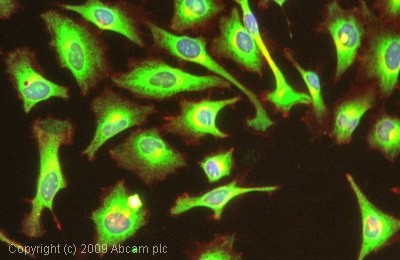 Immunocytochemistry/ Immunofluorescence - Anti-GRP78 BiP antibody (ab32618)ICC/IF image of ab32618 stained HeLa cells. The cells were 100% methanol fixed (5 min) and then incubated in 1%BSA / 10% normal goat serum / 0.3M glycine in 0.1% PBS-Tween for 1h to permeabilise the cells and block non-specific protein-protein interactions. The cells were then incubated with the antibody (ab32618, 1µg/ml) overnight at +4°C. The secondary antibody (green) was Alexa Fluor® 488 goat anti-rabbit IgG (H+L) used at a 1/1000 dilution for 1h. Alexa Fluor® 594 WGA was used to label plasma membranes (red) at a 1/200 dilution for 1h. DAPI was used to stain the cell nuclei (blue) at a concentration of 1.43µM.
-
 Immunohistochemistry (Formalin/PFA-fixed paraffin-embedded sections) - Anti-GRP78 BiP antibody (ab32618)This image shows human breast carcinoma stained with ab32618 diluted 1/100.
 -
 Immunohistochemistry (Formalin/PFA-fixed paraffin-embedded sections) - Anti-GRP78 BiP antibody (ab32618)Ab32618 staining Human normal liver parenchyma. Staining is localised to endoplasmic reticulum compartment.
Left panel: with primary antibody at 2 ug/ml. Right panel: isotype control.
Sections were stained using an automated system DAKO Autostainer Plus , at room temperature: sections were rehydrated and antigen retrieved with the Dako 3 in 1 AR buffers citrate pH6.0 in a DAKO PT Link. Slides were peroxidase blocked in 3% H2O2 in methanol for 10 mins. They were then blocked with Dako Protein block for 10 minutes (containing casein 0.25% in PBS) then incubated with primary antibody for 20 min and detected with Dako envision flex amplification kit for 30 minutes. Colorimetric detection was completed with Diaminobenzidine for 5 minutes. Slides were counterstained with Haematoxylin and coverslipped under DePeX. Please note that for manual staining we recommend to optimize the primary antibody concentration and incubation time (overnight incubation), and amplification may be requi


