Anti-GFP antibody (ab6556)
Key features and details
- Rabbit polyclonal to GFP
- Suitable for: Electron Microscopy, IHC-P, ICC/IF
- Reacts with: Human
- Isotype: IgG
Overview
-
Product name
Anti-GFP antibody
See all GFP primary antibodies -
Description
Rabbit polyclonal to GFP -
Host species
Rabbit -
Specificity
This antibody is reactive against all variants of Aequorea victoria GFP such as S65T-GFP, RS-GFP, YFP, CFP, RFP and EGFP.
-
Tested Applications & Species
See all applications and species dataApplication Species IHC-P Human -
Immunogen
Recombinant full length protein corresponding to GFP.
Database link: P42212 -
General notes
Please note that a mistake was made in reference 4 (Mesaeli et.al., J. Cell. Biol. 1999 Mar 8;144(5):857-68). The antibody used for immunohistochemistry on paraformaldehyde fixed tissues was the crude serum version of this antibody (Abcam ab290) and not Clontech's monoclonal as stated. This product is supplied in 25% glycerol. During freezeing and thawing some phase separation might occur - Please ensure that the solution is mixed thoroughly but GENTLY before use.
This antibody (ab6556) is the purifed version of our best-selling rabbit polyclonal to GFP (ab290). It has been developed specifically for use in applications requiring a high titre and specificity with minimum background such as immuno-electron microscopy.
This anti-GFP antibody recognizes the enhanced form of GFP as well.
Abcam recommended secondaries - Goat Anti-Rabbit HRP (ab205718) and Goat Anti-Rabbit Alexa Fluor® 488 (ab150077).
See other anti-rabbit secondary antibodies that can be used with this antibody.
Properties
-
Form
Liquid -
Storage instructions
Shipped at 4°C. Store at +4°C short term (1-2 weeks). Upon delivery aliquot. Store at -20°C or -80°C. Avoid freeze / thaw cycle. -
Storage buffer
pH: 7.40
Constituents: 0.79% Tris HCl, 25% Glycerol -
 Concentration information loading...
 -
Purity
Immunogen affinity purified -
Purification notes
This antibody is an affinity purified rabbit anti-GFP antibody purified on an affinity chromatography column made with highly purified recombinant GFP. -
Clonality
Polyclonal -
Isotype
IgG -
Research areas
Images
-
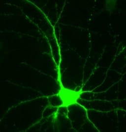
This image shows a single primary hippocampal neuron from a primary culture overexpressing GFP stained with ab6556 at a dilution of 1/2000. This picture was kindly supplied as part of the review submitted by one of our customers.
-
This image shows IF using GFP-expressing glial cells (green) transplanted into lesioned rat spinal cord. This was detected using ab6556 anti-GFP antibody and a FITC conjugated secondary antibody. Axons are labelled red by an antibody to neurofilament-200 and a rhodamine secondary. ab6556 reveals the morphology of the transplanted cells to such an extent that their close interactions with axons are obvious. The top picture shows an optical section from a confocal microscope scan showing how a GFP cell wraps around a branched axon travelling longitudinally. The bottom picture consists of an optical section from another confocal scan showing a GFP cell enveloping an axon in the transverse plane. Review by Andrew Toft submitted 19 May 2004.
-
Specific labeling of a Trk-GFP fusion protein being synthesized on ER in sympathetic neurons infected with an adenovirus carrying the construct. The gold is associated with the ER membranes. This was done using a 1/5000 dilution of affinity purified antibody (ab6556). The tissue section was fixed and embedded using durcupan resin.


