Anti-DDX5 antibody (ab21696)
")
Key features and details
- Rabbit polyclonal to DDX5
- Suitable for: ICC/IF, WB
- Knockout validated
- Reacts with: Mouse, Rat, Human
- Isotype: IgG
Overview
-
Product name
Anti-DDX5 antibody
See all DDX5 primary antibodies -
Description
Rabbit polyclonal to DDX5 -
Host species
Rabbit -
Tested Applications & Species
See all applications and species dataApplication Species ICC/IF HumanWB MouseHuman
-
Immunogen
Synthetic peptide corresponding to Human DDX5 aa 600 to the C-terminus (C terminal) conjugated to keyhole limpet haemocyanin.
(Peptide available asab22412) -
Positive control
- This antibody gave a positive signal in the following lysates: Hela nuclear lysate MEF1 (Mouse embryonic fibroblast cell line) Whole Cell Lysate Testis (Mouse) Tissue Lysate - normal tissue PC12 (Rat adrenal pheochromocytoma cell line) Whole Cell Lysate
Properties
-
Form
Liquid -
Storage instructions
Shipped at 4°C. Store at +4°C short term (1-2 weeks). Upon delivery aliquot. Store at -20°C or -80°C. Avoid freeze / thaw cycle. -
Storage buffer
pH: 7.40
Preservative: 0.02% Sodium azide
Constituent: PBS
Batches of this product that have a concentration Concentration information loading...
Purity
Immunogen affinity purifiedClonality
PolyclonalIsotype
IgGResearch areas
Associated products
-
Compatible Secondaries
-
Isotype control
Applications
The Abpromise guarantee
Our Abpromise guarantee covers the use of ab21696 in the following tested applications.
The application notes include recommended starting dilutions; optimal dilutions/concentrations should be determined by the end user.
GuaranteedTested applications are guaranteed to work and covered by our Abpromise guarantee.
PredictedPredicted to work for this combination of applications and species but not guaranteed.
IncompatibleDoes not work for this combination of applications and species.
Application Species ICC/IF HumanWB MouseHumanApplication Abreviews Notes ICC/IF Use a concentration of 1 µg/ml.WB (3) Use a concentration of 1 µg/ml. Detects a band of approximately 69 kDa (predicted molecular weight: 69 kDa).Notes ICC/IF
Use a concentration of 1 µg/ml.WB
Use a concentration of 1 µg/ml. Detects a band of approximately 69 kDa (predicted molecular weight: 69 kDa).Target
-
Function
RNA-dependent ATPase activity. The rate of ATP hydrolysis is highly stimulated by single-stranded RNA. May be involved in pre-mRNA splicing. -
Sequence similarities
Belongs to the DEAD box helicase family. DDX5/DBP2 subfamily.
Contains 1 helicase ATP-binding domain.
Contains 1 helicase C-terminal domain. -
Post-translational
modificationsArg-502 is dimethylated, probably to asymmetric dimethylarginine. -
Cellular localization
Nucleus > nucleolus. - Information by UniProt
-
Database links
- Entrez Gene: 1655 Human
- Entrez Gene: 13207 Mouse
- Entrez Gene: 287765 Rat
- Omim: 180630 Human
- SwissProt: P17844 Human
- SwissProt: Q61656 Mouse
- Unigene: 279806 Human
- Unigene: 220038 Mouse
see all -
Alternative names
- ATP dependent RNA helicase DDX5 antibody
- DDX 5 antibody
- Ddx5 antibody
see all
Images
-
Western blot - Anti-DDX5 antibody (ab21696)
Lane 1: Wild-type HAP1 whole cell lysate (20 µg)
Lane 2: DDX5 knockout HAP1 whole cell lysate (20 µg)
Lane 3: HeLa whole cell lysate (20 µg)
Lane 4: MCF7 whole cell lysate (20 µg)Lanes 1 - 4: Merged signal (red and green). Green - ab21696 observed at 69 kDa. Red - loading control, ab9484, observed at 37 kDa.
ab21696 was shown to specifically react with DDX5 in wild-type HAP1 cells as signal was lost in DDX5 knockout cells. Wild-type and DDX5 knockout samples were subjected to SDS-PAGE. Ab21696 and ab9484 (Mouse anti-GAPDH loading control) were incubated overnight at 4°C at 1 μg/ml and 1/20000 dilution respectively. Blots were developed with Goat anti-Rabbit IgG H&L (IRDye® 800CW) preabsorbed ab216773 and Goat anti-Mouse IgG H&L (IRDye® 680RD) preabsorbed ab216776 secondary antibodies at 1/20000 dilution for 1 hour at room temperature before imaging.
-
 Western blot - Anti-DDX5 antibody (ab21696)
ab21696 recognizes a clear, strong band at ~ 69kDa corresponding to DDX5. A number of other, non specific bands are weakly recognized by the antibody. All bands are quenched by the addition of the blocking peptide.
-
 Immunocytochemistry/ Immunofluorescence - Anti-DDX5 antibody (ab21696)
ICC/IF image of ab21696 stained human HeLa cells. The cells were PFA fixed (3.7% PFA, 10 min) and incubated with the antibody (ab21696, 1µg/ml) for 1h at room temperature. The secondary antibody (green) was Alexa Fluor® 488 goat anti-rabbit IgG (H+L) used at a 1/1000 dilution for 1h. Image-iTTM FX Signal Enhancer was used as the primary blocking agent, 5% BSA (in TBS-T) was used for all other blocking steps. DAPI was used to stain the cell nuclei (blue). Alexa Fluor® 594 WGA was used to label plasma membranes (red).
ab21696 localises to the nucleus as expected. We believe the small amount of signal seen skirting the nucleus is due to inefficient fixation of the sample.
-
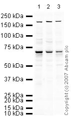 Western blot - Anti-DDX5 antibody (ab21696)All lanes : Anti-DDX5 antibody (ab21696) at 1 µg/ml
Lane 1 : MEF1 (Mouse embryonic fibroblast cell line) Whole Cell Lysate
Lane 2 : Testis (Mouse) Tissue Lysate - normal tissue
Lane 3 : PC12 (Rat adrenal pheochromocytoma cell line) Whole Cell Lysate
Lysates/proteins at 10 µg per lane.
Secondary
All lanes : IRDye 680 Conjugated Goat Anti-Rabbit IgG (H+L) at 1/10000 dilution
Performed under reducing conditions.
Predicted band size: 69 kDa
Observed band size: 70 kDa why is the actual band size different from the predicted?
Additional bands at: 170 kDa. We are unsure as to the identity of these extra bands.
Protocols
References (18)
ab21696 has been referenced in 18 publications.
- Legrand JMD et al. DDX5 plays essential transcriptional and post-transcriptional roles in the maintenance and function of spermatogonia. Nat Commun 10:2278 (2019). PubMed: 31123254
- Fu K et al. Biological and RNA regulatory function of MOV10 in mammalian germ cells. BMC Biol 17:39 (2019). PubMed: 31088452
- Chao TC et al. The Long Noncoding RNA HEAL Regulates HIV-1 Replication through Epigenetic Regulation of the HIV-1 Promoter. mBio 10:N/A (2019). PubMed: 31551335
- Lee YJ et al. Coordinate regulation of alternative pre-mRNA splicing events by the human RNA chaperone proteins hnRNPA1 and DDX5. Genes Dev 32:1060-1074 (2018). PubMed: 30042133
- Rajgor D et al. Identification of novel nesprin-1 binding partners and cytoplasmic matrin-3 in processing bodies. Mol Biol Cell 27:3894-3902 (2016). WB ; Human . PubMed: 27733621
Images
-
Western blot - Anti-DDX5 antibody (ab21696)
Lane 1: Wild-type HAP1 whole cell lysate (20 µg)
Lane 2: DDX5 knockout HAP1 whole cell lysate (20 µg)
Lane 3: HeLa whole cell lysate (20 µg)
Lane 4: MCF7 whole cell lysate (20 µg)Lanes 1 - 4: Merged signal (red and green). Green - ab21696 observed at 69 kDa. Red - loading control, ab9484, observed at 37 kDa.
ab21696 was shown to specifically react with DDX5 in wild-type HAP1 cells as signal was lost in DDX5 knockout cells. Wild-type and DDX5 knockout samples were subjected to SDS-PAGE. Ab21696 and ab9484 (Mouse anti-GAPDH loading control) were incubated overnight at 4°C at 1 μg/ml and 1/20000 dilution respectively. Blots were developed with Goat anti-Rabbit IgG H&L (IRDye® 800CW) preabsorbed ab216773 and Goat anti-Mouse IgG H&L (IRDye® 680RD) preabsorbed ab216776 secondary antibodies at 1/20000 dilution for 1 hour at room temperature before imaging.
-
Western blot - Anti-DDX5 antibody (ab21696)
ab21696 recognizes a clear, strong band at ~ 69kDa corresponding to DDX5. A number of other, non specific bands are weakly recognized by the antibody. All bands are quenched by the addition of the blocking peptide.
-
Immunocytochemistry/ Immunofluorescence - Anti-DDX5 antibody (ab21696)
ICC/IF image of ab21696 stained human HeLa cells. The cells were PFA fixed (3.7% PFA, 10 min) and incubated with the antibody (ab21696, 1µg/ml) for 1h at room temperature. The secondary antibody (green) was Alexa Fluor® 488 goat anti-rabbit IgG (H+L) used at a 1/1000 dilution for 1h. Image-iTTM FX Signal Enhancer was used as the primary blocking agent, 5% BSA (in TBS-T) was used for all other blocking steps. DAPI was used to stain the cell nuclei (blue). Alexa Fluor® 594 WGA was used to label plasma membranes (red).
ab21696 localises to the nucleus as expected. We believe the small amount of signal seen skirting the nucleus is due to inefficient fixation of the sample.
-
Western blot - Anti-DDX5 antibody (ab21696)All lanes : Anti-DDX5 antibody (ab21696) at 1 µg/ml
Lane 1 : MEF1 (Mouse embryonic fibroblast cell line) Whole Cell Lysate
Lane 2 : Testis (Mouse) Tissue Lysate - normal tissue
Lane 3 : PC12 (Rat adrenal pheochromocytoma cell line) Whole Cell Lysate
Lysates/proteins at 10 µg per lane.
Secondary
All lanes : IRDye 680 Conjugated Goat Anti-Rabbit IgG (H+L) at 1/10000 dilution
Performed under reducing conditions.
Predicted band size: 69 kDa
Observed band size: 70 kDa why is the actual band size different from the predicted?
Additional bands at: 170 kDa. We are unsure as to the identity of these extra bands.


