Anti-COX IV antibody - Mitochondrial Loading Control (ab16056)
Key features and details
- Rabbit polyclonal to COX IV - Mitochondrial Loading Control
- Suitable for: WB, ICC/IF, IHC-P
- Reacts with: Mouse, Rat, Human
- Isotype: IgG
Overview
-
Product name
Anti-COX IV antibody - Mitochondrial Loading Control
See all COX IV primary antibodies -
Description
Rabbit polyclonal to COX IV - Mitochondrial Loading Control -
Host species
Rabbit -
Tested Applications & Species
See all applications and species dataApplication Species ICC/IF HumanIHC-P HumanWB MouseHuman -
Immunogen
Synthetic peptide corresponding to Human COX IV aa 150 to the C-terminus (C terminal) conjugated to keyhole limpet haemocyanin.
(Peptide available asab16381) -
General notes
This antibody makes an effective loading control for mitochondria. COX IV is generally expressed at a consistent high level. However, be aware that many proteins run at the same 16kD size as COX IV - our VDAC1 / Porin antibody makes a good alternative mitochondrial loading control for proteins of this size. Some caution is required when using this antibody as a loading control as COXIV expression can vary under some manipulations. An alternative mitochondrial loading control is Mouse monoclonal to COX IV antibody [20E8] (ab14744).
Images
-
All lanes : Anti-COX IV antibody - Mitochondrial Loading Control (ab16056) at 1 µg/ml
Lane 1 : Human skeletal muscle tissue lysate
Lane 2 : Rat skeletal muscle tissue
Lysates/proteins at 10 µg per lane.
Secondary
All lanes : Goat polyclonal to Rabbit IgG - H&L - Pre-Adsorbed (HRP) at 1/50000 dilution
Predicted band size: 17 kDa
Observed band size: 17 kDaBlocking buffer: 2% BSA
Gel type: MES
Exposure Time: 1 minute
-
Lane 1 : Anti-COX IV antibody - Mitochondrial Loading Control (ab16056) at 1 µg/ml (Human skeletal muscle tissue lysate)
Lanes 2-3 : Anti-COX IV antibody - Mitochondrial Loading Control (ab16056) at 1 µg/ml
Lane 2 : Human heart tissue lysate
Lane 3 : Mouse skeletal muscle tissue lysate
Lysates/proteins at 10 µg per lane.
Secondary
All lanes : Goat polyclonal to Rabbit IgG - H&L - Pre-Adsorbed (HRP) at 1/50000 dilution
Predicted band size: 17 kDa
Observed band size: 17 kDaBlocking buffer: 2% BSA
Gel type: MES
Exposure Time: 1 minute
-
All lanes : Anti-COX IV antibody - Mitochondrial Loading Control (ab16056) at 1 µg/ml
Lane 1 : Human brain tissue lysate - total protein (ab29466)
Lane 2 : Human liver tissue lysate - total protein (ab29889)
Lane 3 : Human heart tissue lysate - total protein (ab29431)
Lane 4 : Human skeletal muscle tissue lysate - total protein (ab29330)
Lysates/proteins at 10 µg per lane.
Secondary
All lanes : Goat Anti-Rabbit IgG H&L (HRP) preadsorbed (ab7090) at 1/3000 dilution
Performed under reducing conditions.
Predicted band size: 17 kDa
-
 Immunohistochemistry (Formalin/PFA-fixed paraffin-embedded sections) - Anti-COX IV antibody - Mitochondrial Loading Control (ab16056) This image is courtesy of an anonymous Abreview
ab16056 staining COX IV in Mouse heart tissue sections by Immunohistochemistry (IHC-P - paraformaldehyde-fixed, paraffin-embedded sections). Tissue was fixed with paraformaldehyde and blocked with 5% serum for 1 hour at 21°C; antigen retrieval was by heat mediation in a citrate buffer. Samples were incubated with primary antibody (1/600 ) for 12 hours at 4°C. A Cy5® donkey anti-rabbit secondary antibody was used as the secondary antibody.
-
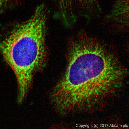
 Immunocytochemistry/ Immunofluorescence - Anti-COX IV antibody - Mitochondrial Loading Control (ab16056)
ab16056 stained in Hela cells. Cells were fixed with 100% methanol (5min) at room temperature and incubated with PBS containing 10% goat serum, 0.3 M glycine, 1% BSA and 0.1% triton for 1h at room temperature to permeabilise the cells and block non-specific protein-protein interactions. The cells were then incubated with the antibody ab16056 at 1µg/ml and ab7291 (Mouse monoclonal [DM1A] to alpha Tubulin - Loading Control) at 1/1000 dilution overnight at +4°C. The secondary antibodies were ab150120 (pseudo-colored red) and ab150081 (colored green) used at 1 ug/ml for 1hour at room temperature. DAPI was used to stain the cell nuclei (colored blue) at a concentration of 1.43µM for 1hour at room temperature
-
All lanes : Anti-COX IV antibody - Mitochondrial Loading Control (ab16056) at 0.38 µg/ml
Lane 1 : HeLa whole cell lysate
Lane 2 : Human skeletal muscle cell lysate
Lane 3 : HeLa whole cell lysate with Human COX IV peptide (ab16381) at 1 µg/ml
Lane 4 : Human skeletal muscle cell lysate with Human COX IV peptide (ab16381) at 1 µg/ml
Lysates/proteins at 20 µg per lane.
Secondary
All lanes : HRP-conjugated goat anti-rabbit IgG at 1/10000 dilution
Predicted band size: 17 kDa
Observed band size: 15 kDa why is the actual band size different from the predicted?
-
 Immunohistochemistry (Formalin/PFA-fixed paraffin-embedded sections) - Anti-COX IV antibody - Mitochondrial Loading Control (ab16056)IHC image of COXIV staining in human liver FFPE section, performed on a BondTM system using the standard protocol F. The section was pre-treated using heat mediated antigen retrieval with sodium citrate buffer (pH6, epitope retrieval solution 1) for 20 mins. The section was then incubated with ab16056, 1µg/ml, for 8 mins at room temperature and detected using an HRP conjugated compact polymer system. DAB was used as the chromogen. The section was then counterstained with haematoxylin and mounted with DPX.
 -
 Immunohistochemistry (Formalin/PFA-fixed paraffin-embedded sections) - Anti-COX IV antibody - Mitochondrial Loading Control (ab16056) This image is courtesy of an anonymous Abreview.
ab16056 staining COX IV in breast tumour tissue sections by Immunohistochemistry (IHC-P - paraformaldehyde-fixed, paraffin-embedded sections). Tissue was fixed with 10% buffered formalin and blocked with 5% serum for 60 minutes at 21°C; antigen retrieval was by heat mediation in a 10mM sodium citrate buffer pH6. Samples were incubated with primary antibody (1/300 in blocking buffer) for 12 hours at 4°C. An Alexa Fluor® 647-conjugated donkey anti-rabbit IgG polyclonal (1/200) was used as the secondary antibody.


