Anti-CD3 antibody (ab828)
")
Key features and details
- Rabbit polyclonal to CD3
- Suitable for: IHC-P, IHC-Fr
- Reacts with: Human
- Isotype: IgG
Overview
-
Product name
Anti-CD3 antibody
See all CD3 primary antibodies -
Description
Rabbit polyclonal to CD3 -
Host species
Rabbit -
Specificity
ab828 is predicted to react with T cell areas in lymphoid tissues derived from various species: human, bovine, porcine, sheep, cat, dog, rat, mouse, Psammomys (sand rat), and chicken. The molecular weight of the polypeptide recognized by the antibody in bovine and swine extracts is 20 kDa.
-
Tested applications
Suitable for: IHC-P, IHC-Frmore details -
Species reactivity
Reacts with: Human -
Immunogen
Synthetic peptide corresponding to Human CD3 conjugated to Bovine Serum Albumin (BSA).
Properties
-
Form
Liquid -
Storage instructions
Shipped at 4°C. Store at +4°C short term (1-2 weeks). Store at -20°C or -80°C. Avoid freeze / thaw cycle. -
Storage buffer
Preservative: 0.05% Sodium azide
Constituent: 1% BSA -
 Concentration information loading...
 -
Purity
Protein A purified -
Clonality
Polyclonal -
Isotype
IgG -
Research areas
Images
-
Immunohistochemistry (Frozen sections) - Anti-CD3 antibody (ab828)
This immunoperoxidase picture shows inflammatory cell infiltration with many T-lymphocytes in the interstitium of human kidneys with congenital nephrotic syndrome of the Finnish type (CNF, NPHS1). Immunoperoxidase staining of frozen sections with ab828 antibody (oval shaped, unstained spaces on the left are glomeruli).
This image was kindly supplied as part of the review submitted by Arvi-Matti-Kuusniemi. -
 Immunohistochemistry (Formalin/PFA-fixed paraffin-embedded sections) - Anti-CD3 antibody (ab828)
Ab828 positively staining formaldehyde fixed human tonsil tissue at 1/50 in conjunction with goat anti rabbit HRP. Detection by DAB.
Pressure cooker heat mediated antigen retrieval was employed in citrate buffer pH 6.0.
This image is courtesy of an Abreview submitted by Melanie Black on 22 Aug 05. We do not have any further information relating to this image.
-
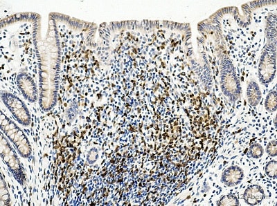
 Immunohistochemistry (Formalin/PFA-fixed paraffin-embedded sections) - Anti-CD3 antibody (ab828) This image is courtesy of an abreview submitted by Carl Hobbs, King's College London, United KingdomIHC-P image of CD3 staining on rat small intestine using ab828 (1:50). The section was deparafinized, rehydrated and subjected to heat mediated antigen retrival using citric acid at pH6. The section was then blocked using 1% BSA at 21°C for 10 mins. The primary antibody was used at 21°C for 2 hours. Goat polyclonal to Rabbit IgG conjugated to biotin was used as secondary antibody (1:250).
 -
 Immunohistochemistry (Formalin/PFA-fixed paraffin-embedded sections) - Anti-CD3 antibody (ab828) This image is courtesy of an abreview submitted by Carl Hobbs, King's College London, United KingdomIHC-P image of CD3 staining on monkey colon using ab828 (1:50). The section was deparafinized, rehydrated and subjected to heat mediated antigen retrival using citric acid at pH6. The section was then blocked using 1% BSA at 21°C for 10 mins. The primary antibody was used at 21°C for 2 hours. Goat polyclonal to Rabbit IgG conjugated to biotin was used as secondary antibody (1:250).
 -
 Immunohistochemistry (Formalin/PFA-fixed paraffin-embedded sections) - Anti-CD3 antibody (ab828) This image is courtesy of an abreview submitted by Carl Hobbs, King's College London, United KingdomIHC-P image of CD3 staining on dog spleen using ab828 (1:50). The section was deparafinized, rehydrated and subjected to heat mediated antigen retrival using citric acid at pH6. The section was then blocked using 1% BSA at 21°C for 10 mins. The primary antibody was used at 21°C for 2 hours. Goat polyclonal to Rabbit IgG conjugated to biotin was used as secondary antibody (1:250).


