Anti-Aurora B antibody (ab2254)
")
Key features and details
- Rabbit polyclonal to Aurora B
- Suitable for: ICC/IF, IHC-P, WB
- Reacts with: Mouse, Rat, Human
- Isotype: IgG
Overview
-
Product name
Anti-Aurora B antibody
See all Aurora B primary antibodies -
Description
Rabbit polyclonal to Aurora B -
Host species
Rabbit -
Tested Applications & Species
See all applications and species dataApplication Species ICC/IF HumanIHC-P HumanWB MouseRatHuman
-
Immunogen
Synthetic peptide within Human Aurora B aa 1-100 conjugated to keyhole limpet haemocyanin. The exact sequence is proprietary.
(Peptide available asab13569)
Images
-
Western blot - Anti-Aurora B antibody (ab2254) Balboula and Schindler PLoS Genet. 2014 Feb 27;10(2):e1004194. doi: 10.1371/journal.pgen.1004194. eCollection 2014 Feb. Fig 1. Reproduced under the Creative Commons license http://creativecommons.org/licenses/by/4.0/
AURKB is expressed in mouse oocytes.
(Panel D) 20 GV-intact oocytes were collected from CF1 mice and micro-injected with the indicated cRNA. Two hours after injection, the oocytes were matured to Met II in vitro (16 h). The total numbers of non-injected control oocytes (Non-inj.) are indicated in parenthesis. Total cellular lysates were probed with the indicated antibody. The panels are images of the same membrane that was stripped and re-probed. The arrows indicate the specific AURKB protein band, and the asterisk indicates a presumed degradation product of AURKB-GFP.
-
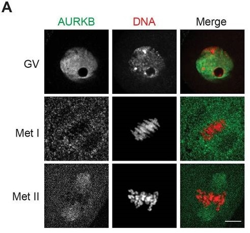 Immunocytochemistry/ Immunofluorescence - Anti-Aurora B antibody (ab2254) Balboula and Schindler PLoS Genet. 2014 Feb 27;10(2):e1004194. doi: 10.1371/journal.pgen.1004194. eCollection 2014 Feb. Fig 1.
AURKB is expressed in mouse oocytes.
(Panel A) GV-intact oocytes were collected from CF1 mice and matured in vitro for 8 h (Met I), or 16 h (Met II), prior to fixation and staining with an anti-AURKB antibody (ab2254).
-
 Immunocytochemistry/ Immunofluorescence - Anti-Aurora B antibody (ab2254) Chopra et al PLoS One. 2016 Apr 20;11(4):e0153818. doi: 10.1371/journal.pone.0153818. eCollection 2016. Fig 6. Reproduced under the Creative Commons license http://creativecommons.org/licenses/by/4.0/
HCT 116 (Human colorectal carcinoma cell line) cells were examined by immunofluorescence confocal microscopy.
Cells were treated with 500 nM AK301 for 16 hours, and then processed for Aurora B (ab2254) and β-tubulin staining (Panel B). The color key and 20 μm bars are shown. Images of representative field is shown with a 20 μm bar. End-labeled DNA is shown in red and DAPI-stained DNA is blue.
Cells cultured on coverslips were fixed with 4% paraformaldehyde at room temperature or 100% ice cold methanol at 4°C and then permeabilized with 0.5% Triton X-100 in PBS. Cells were blocked in 5% serum (in PBS) and then incubated with primary antibody (in 5% serum) on shaker for 1 h at room temperature.
-
 Western blot - Anti-Aurora B antibody (ab2254)All lanes : Anti-Aurora B antibody (ab2254) at 1 µg/ml
Lane 1 : HeLa cell lysate
Lane 2 : HeLa nocodozole treated cell lysate
Lane 3 : NIH3T3 cell lysate
Lane 4 : PC12 cell lysate
Lysates/proteins at 10 µg per lane.
Secondary
All lanes : Goat Anti-Rabbit IgG H&L (HRP) (ab97051) at 1/50000 dilution
Performed under reducing conditions.
Predicted band size: 39 kDaBlocked with 2% BSA.
-
 Immunocytochemistry/ Immunofluorescence - Anti-Aurora B antibody (ab2254)
Immunofluorescence in human cells using Rabbit polyclonal to Aurora B (red), DAPI (blue) and CREST serum (binds to centromeres)(green).
(a) HeLa cells - transition from interphase (left) through mitosis
(b) RPE-1 cells - as in (a)
(c) HeLa cells - interphase
(d) RPE-1 cells - interphase -
 Immunocytochemistry/ Immunofluorescence - Anti-Aurora B antibody (ab2254)
ab2254 stained in Hela cells. Cells were fixed with 4% paraformaldehyde (10min) at room temperature and incubated with PBS containing 10% goat serum, 0.3 M glycine, 1% BSA and 0.1% triton for 1h at room temperature to permeabilise the cells and block non-specific protein-protein interactions. The cells were then incubated with the antibody ab2254 at 1µg/ml and ab7291 (Mouse monoclonal [DM1A] to alpha Tubulin - Loading Control) at 1/1000 dilution overnight at +4°C. The secondary antibodies were ab150120 (pseudo-colored red) and ab150081 (colored green) used at 1 ug/ml for 1hour at room temperature. DAPI was used to stain the cell nuclei (colored blue) at a concentration of 1.43µM for 1hour at room temperature.
-
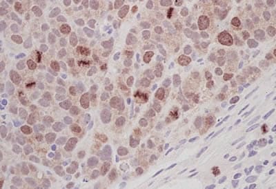
 Immunohistochemistry (Formalin/PFA-fixed paraffin-embedded sections) - Anti-Aurora B antibody (ab2254)
IHC image of Aurora B staining in Human Lymph node Hodgkins disease formalin fixed paraffin embedded tissue section*, performed on a Leica Bond™ system using the standard protocol F. The section was pre-treated using heat mediated antigen retrieval with sodium citrate buffer (pH6, epitope retrieval solution 1) for 20 mins. The section was then incubated with ab2254, 5µg/ml, for 15 mins at room temperature and detected using an HRP conjugated compact polymer system. DAB was used as the chromogen. The section was then counterstained with haematoxylin and mounted with DPX.
For other IHC staining systems (automated and non-automated) customers should optimize variable parameters such as antigen retrieval conditions, primary antibody concentration and antibody incubation times.
*Tissue obtained from the Human Research Tissue Bank, supported by the NIHR Cambridge Biomedical Research Centre
-
 Immunocytochemistry/ Immunofluorescence - Anti-Aurora B antibody (ab2254) This image is courtesy of an Abreview from Lux Fatimathas.
ab2254 staining human A431 (epithelial) cells by ICC/IF. The sample was fixed in paraformaldehyde and permeabilized by incubation with 0.1% Triton X100. 1% BSA was used as the blocking agent prior to a 1 hour incubation with the primary antibody, diluted 1/1000 with 1% BSA made up in PBS. An Alexa Fluor® 647 conjugated Donkey anti-Rabbit IgG (H+L) antibody was used as the secondary. Blocking and antibody incubation steps were carried out at room temperature.
In this set of images, the tubulin is stained green, Aurora B in pink and DNA in blue.
-
 Immunohistochemistry (Formalin/PFA-fixed paraffin-embedded sections) - Anti-Aurora B antibody (ab2254)
Rabbit polyclonal to Aurora B (ab2254) used to stain SW620 human tumour xenografts (in mouse).
The sections were microwave pretreated in citrate buffer (pH 6.0) for 5 mins high then 5 mins simmer (800W conventional microwave). Slides were then incubated for 1 hour with the Aurora B primary antibody diluted 1/200 in TBS, then visualised using DAB, after application of an appropriate secondary. -
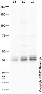 Western blot - Anti-Aurora B antibody (ab2254)All lanes : Anti-Aurora B antibody (ab2254) at 1 µg/ml
Lane 1 : HeLa Whole Cell Lysate
Lane 2 : HeLa Nuclear Lysate
Lane 3 : Jurkat Whole Cell Lysate
Lysates/proteins at 20 µg per lane.
Secondary
All lanes : Goat Anti-Rabbit IgG H&L (HRP) (ab97051) at 1/10000 dilution
Developed using the ECL technique.
Performed under reducing conditions.
Predicted band size: 39 kDa
Observed band size: 39 kDa
Additional bands at: 37 kDa (possible isoform)
Exposure time: 150 seconds
-
 Western blot - Anti-Aurora B antibody (ab2254)Anti-Aurora B antibody (ab2254) at 1/2000 dilution +
Recombinant human Aurora B protein (ab51435) at 0.1 µg
Secondary
Goat Anti-Rabbit IgG H&L (HRP) preadsorbed (ab97080) at 1/5000 dilution
Developed using the ECL technique.
Performed under reducing conditions.
Predicted band size: 39 kDa
Exposure time: 30 seconds


