Recombinant Human Activin Receptor Type IA protein (Fc Chimera) (ab83922)
Key features and details
- Expression system: HEK 293 cells
- Purity: > 95% SDS-PAGE
- Suitable for: SDS-PAGE
-
Product name
Recombinant Human Activin Receptor Type IA protein (Fc Chimera)
See all Activin Receptor Type IA proteins and peptides -
Purity
> 95 % SDS-PAGE. -
Expression system
HEK 293 cells -
Accession
-
Protein length
Protein fragment -
Animal free
No -
Nature
Recombinant -
-
Species
Human -
Sequence
Theoretical Sequence MEDEKPKVNPKLYMCVCEGLSCGNEDHCEGQQCFSSLSINDGFHVYQKGC FQVYEQGKMTCKTPPSPGQAVECCQGDWCNRNITAQLPTKGKSFPGTQNF HLEGSSNTKVDKKVEPKSCDKTHTCPPCPAPELLGGPSVFLFPPKPKDTL MISRTPEVTCVVVDVSHEDPEVKFNWYVDGVEVHNAKTKPREEQYNSTYR VVSVLTVLHQDWLNGKEYKCKVSNKALPAPIEKTISKAKGQPREPQVYTL PPSRDELTKNQVSLTCLVKGFYPSDIAVEWESNGQPENNYKTTPPVLDSD GSFFLYSKLTVDKSRWQQGNVFSCSVMHEALHNHYTQKSLSLSPGK -
Amino acids
1 to 123 -
Additional sequence information
Fused with the Fc region of Human IgG1 at the C-terminus.
-
Preparation and Storage
- Activin A receptor type I
- Activin A receptor type II like kinase 2
- Activin receptor type I
Images
-
Lane 1 – MW markers; Lane 2 – ab83922; Lane 3 – ab83922 treated with PNGase F to remove potential N-linked glycans; Lane 4 – ab83922 treated with a glycosidase cocktail to remove potential N- and O-linked glycans. Approximately 5 μg of protein was loaded per lane; Gel was stained using Coomassie.
Drop in MW after treatment with PNGase F indicates the presence of N-linked glycans. Additional bands in lane 3 and lane 4 are glycosidase enzymes. -
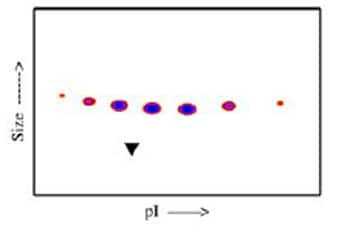
A sample of ab83922 without carrier protein was reduced and alkylated and focused on a 3-10 IPG strip then run on a 4-20% Tris-HCl 2D gel. Approximately 40 μg of protein was loaded; Gel was stained using Deep Purple™. The spot train indicates the presence of multiple glycoforms of ab83922. Spots within the spot train were cut from the gel and identified as Activin Receptor Type IA (Fc Chimera) by protein mass fingerprinting.
-
Post-translational modifications result in protein heterogeneity. The densitometry scan demonstrates the purified human cell expressed protein exists in multiple glycoforms, which differ according to their level of post-translational modification.
The triangle indicates theoretical pI and MW of the protein.


