Mouse E-Cadherin Matched Antibody Pair Kit (ab216783)
Key features and details
- Unlabeled capture antibody, biotin-labeled detection antibody and calibrated protein standard
- For economical ELISA and ELISA-based assay development
- Reacts with: Mouse
- Range: 31.25 pg/ml - 2000 pg/ml
Overview
-
Product name
Mouse E-Cadherin Matched Antibody Pair Kit
See all E Cadherin kits -
Detection method
Colorimetric -
Assay type
ELISA set -
Sensitivity
7.2 pg/ml -
Range
31.25 pg/ml - 2000 pg/ml -
Species reactivity
Reacts with: Mouse -
Product overview
Mouse E-Cadherin Matched Antibody Pair Kits include a capture and a biotinylated detector antibody pair, along with a calibrated protein standard, suitable for sandwich ELISA. The Matched Antibody Pair Kit can be used to quantify native and recombinant mouse E-Cadherin.
Optimization of the kit reagents to sample type, immunoassay format or instrumentation may be required. Guidelines for use of this kit in a standard 96-well microplate sandwich ELISA using HRP/TMB system of colorimetric detection is described in this assay procedure for the purposes of quantification.
Protocol information and tips on the use of the Matched Antibody Pair kits for sandwich ELISA can be found on our website. An accessory pack can be purchased which includes buffer reagents required to perform 10 x 96-well plate sandwich ELISAs (ab210905).
For additional information on the performance of the antibody pair used in this kit, please see our equivalent SimpleStep ELISA kit ab197751. Please note that while the antibody pair is the same provided in the corresponding SimpleStep ELISA Kit, due to differences in their formulation, this antibody pair cannot be used with the consumables provided with our SimpleStep ELISA Kits.
-
Tested applications
Suitable for: ELISAmore details -
Platform
Reagents
Properties
-
Storage instructions
Store at -20°C. Please refer to protocols. -
Components 10 x 96 tests 5 x 96 tests Mouse E-Cadherin Capture Antibody 2 x 50µg 1 x 50µg Mouse E-Cadherin Detector Antibody 2 x 12.5µg 1 x 12.5µg Mouse E-Cadherin Lyophilized Protein 2 vials 1 vial -
Research areas
-
Function
Cadherins are calcium-dependent cell adhesion proteins. They preferentially interact with themselves in a homophilic manner in connecting cells; cadherins may thus contribute to the sorting of heterogeneous cell types. CDH1 is involved in mechanisms regulating cell-cell adhesions, mobility and proliferation of epithelial cells. Has a potent invasive suppressor role. It is a ligand for integrin alpha-E/beta-7.
E-Cad/CTF2 promotes non-amyloidogenic degradation of Abeta precursors. Has a strong inhibitory effect on APP C99 and C83 production. -
Tissue specificity
Non-neural epithelial tissues. -
Involvement in disease
Defects in CDH1 are the cause of hereditary diffuse gastric cancer (HDGC) [MIM:137215]. An autosomal dominant cancer predisposition syndrome with increased susceptibility to diffuse gastric cancer. Diffuse gastric cancer is a malignant disease characterized by poorly differentiated infiltrating lesions resulting in thickening of the stomach. Malignant tumors start in the stomach, can spread to the esophagus or the small intestine, and can extend through the stomach wall to nearby lymph nodes and organs. It also can metastasize to other parts of the body. Note=Heterozygous germline mutations CDH1 are responsible for familial cases of diffuse gastric cancer. Somatic mutations in the has also been found in patients with sporadic diffuse gastric cancer and lobular breast cancer.
Defects in CDH1 are a cause of susceptibility to endometrial cancer (ENDMC) [MIM:608089].
Defects in CDH1 are a cause of susceptibility to ovarian cancer (OC) [MIM:167000]. Ovarian cancer common malignancy originating from ovarian tissue. Although many histologic types of ovarian neoplasms have been described, epithelial ovarian carcinoma is the most common form. Ovarian cancers are often asymptomatic and the recognized signs and symptoms, even of late-stage disease, are vague. Consequently, most patients are diagnosed with advanced disease. -
Sequence similarities
Contains 5 cadherin domains. -
Post-translational
modificationsDuring apoptosis or with calcium influx, cleaved by a membrane-bound metalloproteinase (ADAM10), PS1/gamma-secretase and caspase-3 to produce fragments of about 38 kDa (E-CAD/CTF1), 33 kDa (E-CAD/CTF2) and 29 kDa (E-CAD/CTF3), respectively. Processing by the metalloproteinase, induced by calcium influx, causes disruption of cell-cell adhesion and the subsequent release of beta-catenin into the cytoplasm. The residual membrane-tethered cleavage product is rapidly degraded via an intracellular proteolytic pathway. Cleavage by caspase-3 releases the cytoplasmic tail resulting in disintegration of the actin microfilament system. The gamma-secretase-mediated cleavage promotes disassembly of adherens junctions. -
Cellular localization
Cell junction. Cell membrane. Endosome. Golgi apparatus > trans-Golgi network. Colocalizes with DLGAP5 at sites of cell-cell contact in intestinal epithelial cells. Anchored to actin microfilaments through association with alpha-, beta- and gamma-catenin. Sequential proteolysis induced by apoptosis or calcium influx, results in translocation from sites of cell-cell contact to the cytoplasm. Colocalizes with RAB11A endosomes during its transport from the Golgi apparatus to the plasma membrane. - Information by UniProt
-
Alternative names
- Arc 1
- CADH1_HUMAN
- Cadherin 1
see all -
Database links
- Entrez Gene: 12550 Mouse
- SwissProt: P09803 Mouse
- Unigene: 35605 Mouse
Images
-
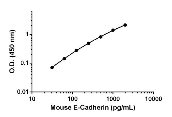
Standard calibration curve. Background subtracted values are graphed.
-
To learn more about the advantages of recombinant antibodies see here.


