Human p53 ELISA Kit (ab171571)
")
Key features and details
- One-wash 90 minute protocol
- Sensitivity: 65 pg/ml
- Range: 0.23 ng/ml - 15 ng/ml
- Sample type: Cell culture extracts
- Detection method: Colorimetric
- Assay type: Sandwich (quantitative)
- Reacts with: Human
Overview
-
Product name
Human p53 ELISA Kit
See all p53 kits -
Detection method
Colorimetric -
Precision
Intra-assay Sample n Mean SD CV% Overall 5 4.3% Inter-assay Sample n Mean SD CV% Overall 3 6.7% -
Sample type
Cell culture extracts -
Assay type
Sandwich (quantitative) -
Sensitivity
65 pg/ml -
Range
0.23 ng/ml - 15 ng/ml -
Recovery
Sample specific recovery Sample type Average % Range Cell culture media 99 93% - 107% Fetal Bovine Serum 93 85% - 104% -
Assay time
1h 30m -
Assay duration
One step assay -
Species reactivity
Reacts with: Human -
Product overview
Human p53 ELISA kit (ab171571) is a single-wash 90 min sandwich ELISA designed for the quantitative measurement of p53 protein in human cell samples. It uses our proprietary SimpleStep ELISA® technology. Quantitate human p53 with 65 pg/ml sensitivity.
SimpleStep ELISA® technology employs capture antibodies conjugated to an affinity tag that is recognized by the monoclonal antibody used to coat our SimpleStep ELISA® plates. This approach to sandwich ELISA allows the formation of the antibody-analyte sandwich complex in a single step, significantly reducing assay time. See the SimpleStep ELISA® protocol summary in the image section for further details. Our SimpleStep ELISA® technology provides several benefits:
-Single-wash protocol reduces assay time to 90 minutes or less
-High sensitivity, specificity and reproducibility from superior antibodies
-Fully validated in biological samples
-96-wells plate breakable into 12 x 8 wells strips
A 384-well SimpleStep ELISA® microplate (ab203359) is available to use as an alternative to the 96-well microplate provided with SimpeStep ELISA® kits. -
Notes
p53 (TP53 gene) acts as a tumor suppressor in many tumor types and induces growth arrest or apoptosis depending on the physiological circumstances and cell type. p53 is involved in cell cycle regulation as a trans-activator that acts to negatively regulate cell division by controlling a set of genes required for this process. One of the activated genes is an inhibitor of cyclin-dependent kinases. p53 mediated apoptosis induction seems to be by stimulation of BAX and FAS antigen expression, or by repression of Bcl-2 expression. p53 is also implicated in Notch signaling crossover
The p53 protein is found in increased amounts in a wide variety of transformed cells. p53 is mutated or inactivated in about 60% of cancers. Four types of cancers account for 80% of tumors occurring in TP53 germline mutation carriers: breast cancers, soft tissue and bone sarcomas, brain tumors (astrocytomas) and adrenocortical carcinomas.
p53 levels are kept low through a continuous degradation of p53. Mdm2 binds to p53, preventing its action and transports it from the nucleus to the cytosol. Mdm2 also acts as ubiquitin ligase and covalently attaches ubiquitin to p53 and thus marks p53 for degradation by the proteasome. The ubiquitin can be cleaved by USP7 (or HAUSP), thereby protecting it from this proteasome dependent degradation. This is one means by which p53 is stabilized in response to oncogenic insults.
Phosphorylation of the N-terminal end of p53, and conformational changes to p53, disrupt Mdm2-binding leading to p53 accumulation. Acetylation of the C-terminal end of p53 exposes the DNA binding domain of p53, allowing it to activate or repress specific genes.
Deacetylase enzymes, such as Sirt1 and Sirt7, can deacetylate p53, leading to an inhibition of apoptosis.
Abcam has not and does not intend to apply for the REACH Authorisation of customers’ uses of products that contain European Authorisation list (Annex XIV) substances.
It is the responsibility of our customers to check the necessity of application of REACH Authorisation, and any other relevant authorisations, for their intended uses. -
Platform
Microplate
Properties
-
Storage instructions
Store at +4°C. Please refer to protocols. -
Components 1 x 96 tests 10X Human p53 Capture Antibody 1 x 600µl 10X Human p53 Detector Antibody 1 x 600µl 10X Wash Buffer PT (ab206977) 1 x 20ml 4X Antibody Diluent EB 1 x 6ml 50X Cell Extraction Enhancer Solution (ab193971) 1 x 1ml 5X Cell Extraction Buffer PTR (ab193970) 1 x 10ml Human p53 Lyophilized Recombinant Protein 2 vials Plate Seals 1 unit Sample Diluent NS (ab193972) 1 x 12ml SimpleStep Pre-Coated 96-Well Microplate (ab206978) 1 unit Stop Solution 1 x 12ml TMB Development Solution 1 x 12ml -
Research areas
-
Function
Acts as a tumor suppressor in many tumor types; induces growth arrest or apoptosis depending on the physiological circumstances and cell type. Involved in cell cycle regulation as a trans-activator that acts to negatively regulate cell division by controlling a set of genes required for this process. One of the activated genes is an inhibitor of cyclin-dependent kinases. Apoptosis induction seems to be mediated either by stimulation of BAX and FAS antigen expression, or by repression of Bcl-2 expression. Implicated in Notch signaling cross-over. Isoform 2 enhances the transactivation activity of isoform 1 from some but not all TP53-inducible promoters. Isoform 4 suppresses transactivation activity and impairs growth suppression mediated by isoform 1. Isoform 7 inhibits isoform 1-mediated apoptosis. -
Tissue specificity
Ubiquitous. Isoforms are expressed in a wide range of normal tissues but in a tissue-dependent manner. Isoform 2 is expressed in most normal tissues but is not detected in brain, lung, prostate, muscle, fetal brain, spinal cord and fetal liver. Isoform 3 is expressed in most normal tissues but is not detected in lung, spleen, testis, fetal brain, spinal cord and fetal liver. Isoform 7 is expressed in most normal tissues but is not detected in prostate, uterus, skeletal muscle and breast. Isoform 8 is detected only in colon, bone marrow, testis, fetal brain and intestine. Isoform 9 is expressed in most normal tissues but is not detected in brain, heart, lung, fetal liver, salivary gland, breast or intestine. -
Involvement in disease
Note=TP53 is found in increased amounts in a wide variety of transformed cells. TP53 is frequently mutated or inactivated in about 60% of cancers. TP53 defects are found in Barrett metaplasia a condition in which the normally stratified squamous epithelium of the lower esophagus is replaced by a metaplastic columnar epithelium. The condition develops as a complication in approximately 10% of patients with chronic gastroesophageal reflux disease and predisposes to the development of esophageal adenocarcinoma.
Defects in TP53 are a cause of esophageal cancer (ESCR) [MIM:133239].
Defects in TP53 are a cause of Li-Fraumeni syndrome (LFS) [MIM:151623]. LFS is an autosomal dominant familial cancer syndrome that in its classic form is defined by the existence of a proband affected by a sarcoma before 45 years with a first degree relative affected by any tumor before 45 years and another first degree relative with any tumor before 45 years or a sarcoma at any age. Other clinical definitions for LFS have been proposed (PubMed:8118819 and PubMed:8718514) and called Li-Fraumeni like syndrome (LFL). In these families affected relatives develop a diverse set of malignancies at unusually early ages. Four types of cancers account for 80% of tumors occurring in TP53 germline mutation carriers: breast cancers, soft tissue and bone sarcomas, brain tumors (astrocytomas) and adrenocortical carcinomas. Less frequent tumors include choroid plexus carcinoma or papilloma before the age of 15, rhabdomyosarcoma before the age of 5, leukemia, Wilms tumor, malignant phyllodes tumor, colorectal and gastric cancers.
Defects in TP53 are involved in head and neck squamous cell carcinomas (HNSCC) [MIM:275355]; also known as squamous cell carcinoma of the head and neck.
Defects in TP53 are a cause of lung cancer (LNCR) [MIM:211980].
Defects in TP53 are a cause of choroid plexus papilloma (CPLPA) [MIM:260500]. Choroid plexus papilloma is a slow-growing benign tumor of the choroid plexus that often invades the leptomeninges. In children it is usually in a lateral ventricle but in adults it is more often in the fourth ventricle. Hydrocephalus is common, either from obstruction or from tumor secretion of cerebrospinal fluid. If it undergoes malignant transformation it is called a choroid plexus carcinoma. Primary choroid plexus tumors are rare and usually occur in early childhood.
Defects in TP53 are a cause of adrenocortical carcinoma (ADCC) [MIM:202300]. ADCC is a rare childhood tumor of the adrenal cortex. It occurs with increased frequency in patients with the Beckwith-Wiedemann syndrome and is a component tumor in Li-Fraumeni syndrome. -
Sequence similarities
Belongs to the p53 family. -
Domain
The nuclear export signal acts as a transcriptional repression domain. The TADI and TADII motifs (residues 17 to 25 and 48 to 56) correspond both to 9aaTAD motifs which are transactivation domains present in a large number of yeast and animal transcription factors. -
Post-translational
modificationsAcetylated. Acetylation of Lys-382 by CREBBP enhances transcriptional activity. Deacetylation of Lys-382 by SIRT1 impairs its ability to induce proapoptotic program and modulate cell senescence.
Phosphorylation on Ser residues mediates transcriptional activation. Phosphorylated by HIPK1 (By similarity). Phosphorylation at Ser-9 by HIPK4 increases repression activity on BIRC5 promoter. Phosphorylated on Thr-18 by VRK1. Phosphorylated on Ser-20 by CHEK2 in response to DNA damage, which prevents ubiquitination by MDM2. Phosphorylated on Thr-55 by TAF1, which promotes MDM2-mediated degradation. Phosphorylated on Ser-46 by HIPK2 upon UV irradiation. Phosphorylation on Ser-46 is required for acetylation by CREBBP. Phosphorylated on Ser-392 following UV but not gamma irradiation. Phosphorylated upon DNA damage, probably by ATM or ATR. Phosphorylated on Ser-15 upon ultraviolet irradiation; which is enhanced by interaction with BANP.
Dephosphorylated by PP2A-PPP2R5C holoenzyme at Thr-55. SV40 small T antigen inhibits the dephosphorylation by the AC form of PP2A.
May be O-glycosylated in the C-terminal basic region. Studied in EB-1 cell line.
Ubiquitinated by MDM2 and SYVN1, which leads to proteasomal degradation. Ubiquitinated by RFWD3, which works in cooperation with MDM2 and may catalyze the formation of short polyubiquitin chains on p53/TP53 that are not targeted to the proteasome. Ubiquitinated by MKRN1 at Lys-291 and Lys-292, which leads to proteasomal degradation. Deubiquitinated by USP10, leading to its stabilization. Ubiquitinated by TRIM24, which leads to proteasomal degradation. Ubiquitination by TOPORS induces degradation. Deubiquitination by USP7, leading to stabilization. Isoform 4 is monoubiquitinated in an MDM2-independent manner.
Monomethylated at Lys-372 by SETD7, leading to stabilization and increased transcriptional activation. Monomethylated at Lys-370 by SMYD2, leading to decreased DNA-binding activity and subsequent transcriptional regulation activity. Lys-372 monomethylation prevents interaction with SMYD2 and subsequent monomethylation at Lys-370. Dimethylated at Lys-373 by EHMT1 and EHMT2. Monomethylated at Lys-382 by SETD8, promoting interaction with L3MBTL1 and leading to repress transcriptional activity. Demethylation of dimethylated Lys-370 by KDM1A prevents interaction with TP53BP1 and represses TP53-mediated transcriptional activation.
Sumoylated by SUMO1. -
Cellular localization
Cytoplasm; Cytoplasm. Nucleus. Nucleus > PML body. Endoplasmic reticulum. Interaction with BANP promotes nuclear localization. Recruited into PML bodies together with CHEK2; Nucleus. Cytoplasm. Localized in both nucleus and cytoplasm in most cells. In some cells, forms foci in the nucleus that are different from nucleoli; Nucleus. Cytoplasm. Localized in the nucleus in most cells but found in the cytoplasm in some cells; Nucleus. Cytoplasm. Localized mainly in the nucleus with minor staining in the cytoplasm; Nucleus. Cytoplasm. Predominantly nuclear but localizes to the cytoplasm when expressed with isoform 4 and Nucleus. Cytoplasm. Predominantly nuclear but translocates to the cytoplasm following cell stress. - Information by UniProt
-
Alternative names
- Antigen NY-CO-13
- BCC7
- Cellular tumor antigen p53
see all -
Database links
- Entrez Gene: 7157 Human
- Omim: 191170 Human
- SwissProt: P04637 Human
- Unigene: 654481 Human
Images
-
Other - Human p53 ELISA Kit (ab171571)
SimpleStep ELISA technology allows the formation of the antibody-antigen complex in one single step, reducing assay time to 90 minutes. Add samples or standards and antibody mix to wells all at once, incubate, wash, and add your final substrate. See protocol for a detailed step-by-step guide.
-
 Example p53 Standard Curve
 -
 Titration of HEK293 extract within the working range of the assay
 -
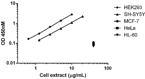 Comparison of p53 expression in different cell lines
Background subtracted data from duplicate measurements are plotted. Note the relative lower expression of p53 in the MCF7, HeLa and HL60 cell lines compared to HEK293 and SH-SY5Y.
-
 Comparison of p53 expression in different cell lines
ELISA (barchart) and western blot (top). Raw OD450 nm data from duplicate measurements for the indicated cells lines is shown (20 µg lysate analyzed). The p53 capture antibody was used to blot the same lysates as analyzed by ELISA (20 µg loaded/lane). The GAPDH blot is included to show the relative loads of each lysate.


