Anti-ATRX antibody (ab97508)
Key features and details
- Rabbit polyclonal to ATRX
- Suitable for: IP, WB, IHC-P, ICC/IF
- Reacts with: Mouse, Rat, Human
- Isotype: IgG
Overview
-
Product name
Anti-ATRX antibody
See all ATRX primary antibodies -
Description
Rabbit polyclonal to ATRX -
Host species
Rabbit -
Tested Applications & Species
See all applications and species dataApplication Species ICC/IF HumanIHC-P HumanIP HumanWB MouseRatHuman -
Immunogen
Recombinant fragment corresponding to Human ATRX aa 2211-2413.
Database link: P46100 -
Positive control
- 293T, HepG2, Raji, NIH-3T3, HeLa A431, H1299 and HeLaS3 cell lines. A549 xenograft.
Properties
-
Form
Liquid -
Storage instructions
Shipped at 4°C. Upon delivery aliquot. Store at -20°C or -80°C. Avoid freeze / thaw cycle. -
Storage buffer
pH: 7.00
Preservative: 0.025% Proclin 300
Constituents: 79% PBS, 20% Glycerol (glycerin, glycerine) -
 Concentration information loading...
 -
Purity
Immunogen affinity purified -
Clonality
Polyclonal -
Isotype
IgG -
Research areas
Images
-
Western blot of (lane1) PC12 (brain)whole cell extracts, (lane 2) Rat 2 (embryo) whole cell extracts, at 30µg per lane, labeling ATRX with ab97508 at 1:1000. A HRP-conjugated anti-rabbit IgG antibody was used to detect the primary antibody.
-
Immunohistochemistry (Formalin/PFA-fixed paraffin-embedded sections) analysis of human breast carcinoma labeling ATRX with ab97508 at 1/500 dilution. Anti-ATRX antibody (ab97508) detects ATRX protein at nucleus on human breast carcinoma tissue sections.
-
All lanes : Anti-ATRX antibody (ab97508) at 1/500 dilution
Lane 1 : 293T whole cell lysate
Lane 2 : HepG2 whole cell lysate
Lane 3 : Raji whole cell lysate
Lysates/proteins at 30 µg per lane.
Predicted band size: 283 kDa
5% SDS-PAGE -
Immunohistochemistry (Formalin/PFA-fixed paraffin-embedded sections) analysis of endometrial carcinoma labeling ATRX with ab97508 at 1/500 dilution. Anti-ATRX antibody (ab97508) detects ATRX protein at nucleus on human endometrial carcinoma tissue sections.
-
Immunohistochemical analysis of paraffin-embedded human lung carcinoma,using ab97508 at 1/500 dilution.
-
 Immunocytochemistry/ Immunofluorescence - Anti-ATRX antibody (ab97508) Image from Heckman, L. D., et al. eLife 3 (2014): e02676. doi: 10.7554/eLife.02676. Fig 4D. Reproduced under the Creative Commons license http://creativecommons.org/licenses/by/4.0/
Immunocytochemistry/ Immunofluorescence analysis of mouse brain labeling ATRX with ab97508 at 1/100 dilution. Left- DAPI (blue), middle - ATRX protein staining (red), right - merge.
-
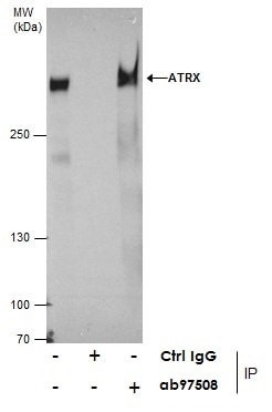
ab97508 at 5µg immunoprecipitating ATRX in 293T whole cell extracts.
Lane 1 (input): 293T whole cell extracts.
Lane 2 (-): Rabbit IgG instead of ab97508 in 293T whole cell extracts.
Lane 3 (+): Anti-ATRX antibody (ab97508) + 293T whole cell extracts. -
Anti-ATRX antibody (ab97508) at 1/1000 dilution + NIH3T3 whole cell extracts at 30 µg
Predicted band size: 283 kDa
-
Immunofluorescence analysis of paraformaldehyde-fixed HeLa cells, using ab97508 at 1/200 dilution. Lower image is merged with DNA probe.
-
Anti-ATRX antibody (ab97508) at 1/1000 dilution + NIH-3T3 whole cell lysate at 30 µg
Predicted band size: 283 kDa
5% SDS-PAGE


