Human Fibronectin ELISA Kit, Fluorescent (ab229398)
Key features and details
- One-wash 90 minute protocol
- Sensitivity: 26 pg/ml
- Range: 31 pg/ml - 32000 pg/ml
- Sample type: Cell culture extracts, Cell culture supernatant, Cit plasma, EDTA Plasma, Milk, Serum, Tissue Extracts
- Detection method: Fluorescent
- Assay type: Sandwich (quantitative)
- Reacts with: Human, Rhesus monkey
Overview
-
Product name
Human Fibronectin ELISA Kit, Fluorescent
See all Fibronectin kits -
Detection method
Fluorescent -
Precision
Intra-assay Sample n Mean SD CV% Serum 3 5% Inter-assay Sample n Mean SD CV% Serum 5 8.3% -
Sample type
Cell culture supernatant, Milk, Serum, Cell culture extracts, Tissue Extracts, EDTA Plasma, Cit plasma -
Assay type
Sandwich (quantitative) -
Sensitivity
26 pg/ml -
Range
31 pg/ml - 32000 pg/ml -
Recovery
Sample specific recovery Sample type Average % Range Cell culture supernatant 102 90% - 117% Milk 102 91% - 111% Serum 93 87% - 102% Cell culture extracts 102 90% - 117% Tissue Extracts 90 80% - 131% EDTA Plasma 86 81% - 94% Cit plasma 99 87% - 120% -
Assay time
1h 30m -
Assay duration
One step assay -
Species reactivity
Reacts with: Human, Rhesus monkey
Does not react with: Cow -
Product overview
Fibronectin in vitro CatchPoint® SimpleStep ELISA® (Enzyme-Linked Immunosorbent Assay) kit is designed for the quantitative measurement of Fibronectin protein in human serum, plasma, milk, cell culture supernatant, and cell and tissue extract samples.
This CatchPoint SimpleStep ELISA kit has been optimized for Molecular Devices Microplate Readers. Click here for a list of recommended Microplate Readers.
If using a Molecular Devices’ plate reader supported by SoftMax® Pro software, a preconfigured protocol for these CatchPoint SimpleStep ELISA Kits is available with all the protocol and analysis settings at www.softmaxpro.org.The CatchPoint® SimpleStep ELISA® employs an affinity tag labeled capture antibody and a reporter conjugated detector antibody which immunocapture the sample analyte in solution. This entire complex (capture antibody/analyte/detector antibody) is in turn immobilized via immunoaffinity of an anti-tag antibody coating the well. To perform the assay, samples or standards are added to the wells, followed by the antibody mix. After incubation, the wells are washed to remove unbound material. CatchPoint HRP Development Solution containing the Stoplight Red Substrate is added. During incubation, the substrate is catalyzed by HRP generating a fluorescent product. Signal is generated proportionally to the amount of bound analyte and the intensity is measured in a fluorescence plater reader at 530/570/590 nm Excitation/Cutoff/Emission.
-
Notes
Fibronectin is a large glycoprotein present in the extracellular matrix and circulating plasma. Fibronectin is important in many cell adhesion and migration related processes, including wound healing, embryogenesis and nerve regeneration. Differential expression of fibronectin is seen in coronary heart disease, glomerulopathy and tumor cell metastasis. The protein contains binding sites for collagen, heparin and fibrin and is a specific ligand for several integrin adhesion receptors. Fibronectin exists as a dimer or multimer.
Abcam has not and does not intend to apply for the REACH Authorisation of customers’ uses of products that contain European Authorisation list (Annex XIV) substances.
It is the responsibility of our customers to check the necessity of application of REACH Authorisation, and any other relevant authorisations, for their intended uses. -
Platform
Pre-coated microplate (12 x 8 well strips)
Properties
-
Storage instructions
Store at +4°C. Please refer to protocols. -
Components 1 x 96 tests 100X Stoplight Red Substrate 1 x 120µl 10X Human Fibronectin Capture Antibody 1 x 600µl 10X Human Fibronectin Detector Antibody 1 x 600µl 10X Wash Buffer PT (ab206977) 1 x 20ml 500X Hydrogen Peroxide (H2O2, 3%) 1 x 50µl 50X Cell Extraction Enhancer Solution (ab193971) 1 x 1ml 5X Cell Extraction Buffer PTR (ab193970) 1 x 10ml Antibody Diluent 4BR 1 x 6ml Human Fibronectin Lyophilized Recombinant Protein 2 vials Plate Seals 1 unit Sample Diluent NS (ab193972) 1 x 50ml SimpleStep Pre-Coated Black 96-Well Microplate 1 unit Stoplight Red Substrate Buffer 1 x 12ml -
Research areas
-
Function
Fibronectins bind cell surfaces and various compounds including collagen, fibrin, heparin, DNA, and actin. Fibronectins are involved in cell adhesion, cell motility, opsonization, wound healing, and maintenance of cell shape. Involved in osteoblast compaction through the fibronectin fibrillogenesis cell-mediated matrix assembly process, essential for osteoblast mineralization. Participates in the regulation of type I collagen deposition by osteoblasts.
Anastellin binds fibronectin and induces fibril formation. This fibronectin polymer, named superfibronectin, exhibits enhanced adhesive properties. Both anastellin and superfibronectin inhibit tumor growth, angiogenesis and metastasis. Anastellin activates p38 MAPK and inhibits lysophospholipid signaling. -
Tissue specificity
Plasma FN (soluble dimeric form) is secreted by hepatocytes. Cellular FN (dimeric or cross-linked multimeric forms), made by fibroblasts, epithelial and other cell types, is deposited as fibrils in the extracellular matrix. Ugl-Y1, Ugl-Y2 and Ugl-Y3 are found in urine. -
Involvement in disease
Glomerulopathy with fibronectin deposits 2 -
Sequence similarities
Contains 12 fibronectin type-I domains.
Contains 2 fibronectin type-II domains.
Contains 16 fibronectin type-III domains. -
Developmental stage
Ugl-Y1, Ugl-Y2 and Ugl-Y3 are present in the urine from 0 to 17 years of age. -
Post-translational
modificationsSulfated.
It is not known whether both or only one of Thr-2064 and Thr-2065 are/is glycosylated.
Forms covalent cross-links mediated by a transglutaminase, such as F13A or TGM2, between a glutamine and the epsilon-amino group of a lysine residue, forming homopolymers and heteropolymers (e.g. fibrinogen-fibronectin, collagen-fibronectin heteropolymers).
Phosphorylated by FAM20C in the extracellular medium.
Proteolytic processing produces the C-terminal NC1 peptide, anastellin. -
Cellular localization
Secreted, extracellular space, extracellular matrix. - Information by UniProt
-
Alternative names
- CIG
- Cold insoluble globulin
- Cold-insoluble globulin
see all -
Database links
- Entrez Gene: 2335 Human
- Omim: 135600 Human
- SwissProt: P02751 Human
- Unigene: 203717 Human
Images
-
SimpleStep ELISA technology allows the formation of the antibody-antigen complex in one single step, reducing assay time to 90 minutes. Add samples or standards and antibody mix to wells all at once, incubate, wash, and add your final substrate. See protocol for a detailed step-by-step guide.
-
Background-subtracted data values (mean +/- SD) are graphed.
-
The concentrations of Fibronectin were measured in duplicates, interpolated from the Fibronectin standard curves and corrected for sample dilution. Undiluted samples are as follows: serum 1/20,000, plasma (citrate) 1/20,000, and plasma (EDTA) 1/40,000. The interpolated dilution factor corrected values are plotted (mean +/- SD, n=2). The mean Fibronectin concentration was determined to be 126.5 µg/mL in serum, 141.5 µg/mL in plasma (citrate), and 232.1 µg/mL in plasma (EDTA).
-
 Interpolated concentrations of native Fibronectin in human breast milk and HepG2 cell culture supernatant (4 days) samples.
The concentrations of Fibronectin were measured in duplicates, interpolated from the Fibronectin standard curves and corrected for sample dilution. Undiluted samples are as follows: milk 1/100, and HepG2 cell culture supernatant 1/2000. The interpolated dilution factor corrected values are plotted (mean +/- SD, n=2). The mean Fibronectin concentration was determined to be 502.9 ng/mL in milk, and 9683 ng/mL in HepG2 cell culture supernatant.
-
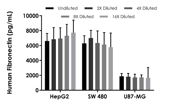
Interpolated concentrations of native Fibronectin in human HepG2 cell extract samples based on a 25 μg/mL extract load, SW 480 cell extract samples based on a 100 μg/mL extract load, and U87-MG cell extract samples based on a 5 μg/mL extract load. The concentrations of Fibronectin were measured in duplicate and interpolated from the Fibronectin standard curve and corrected for sample dilution. The interpolated dilution factor corrected values are plotted (mean +/- SD, n=2). The mean Fibronectin concentration was determined to be 7085 pg/mL in HepG2 cell extract, 6314 pg/mL in SW 480 cell extract, and 1749 pg/mL in U87-MG cell extract.
-
Interpolated concentrations of native Fibronectin in human liver tissue extract based on a 100 μg/mL extract load, heart tissue extract based on a 100 μg/mL extract load, skeletal muscle based on a 100 μg/mL extract load, colon tissue extract based on a 5 μg/mL extract load, and placenta tissue extract based on a 1.25 μg/mL extract load. The concentrations of Fibronectin were measured in duplicate and interpolated from the Fibronectin standard curve and corrected for sample dilution. The interpolated dilution factor corrected values are plotted (mean +/- SD, n=2). The mean Fibronectin concentration was determined to be 7634 pg/mL in liver tissue extract, 4910 pg/mL in heart tissue extract, 4278 pg/mL in skeletal muscle tissue extract, 2684 pg/mL in colon tissue extract, and 1224 pg/mL in placenta tissue extract.
-
Interpolated dilution factor corrected values are plotted (mean +/- SD, n=2). The mean Fibronectin concentration was determined to be 146.2 µg/mL with a range of 35.99 – 319.2 µg/mL.
-
The concentrations of Fibronectin were measured in three different dilutions in duplicate and interpolated from the Fibronectin standard curve and corrected for sample dilution. The interpolated dilution factor corrected values are plotted in ng of Fibronectin per mg of extract (mean +/- SD, n=3). Fibronectin concentration was determined to be 271.8 ng/mg in HepG2 cell extract, 65.52 ng/mg in SW 480 cell extract, 359.6 ng/mg in U87-MG cell extract, 75.22 ng/mg in liver tissue extract, 47.20 ng/mg in heart tissue extract, 43.52 ng/mg in skeletal muscle tissue extract, 557.9 ng/mg in colon tissue extract, and 998.8 ng/mg in placenta tissue extract samples.
-
To learn more about the advantages of recombinant antibodies see here.


