EpCAM (EPR20532-225, AUA1, EPR20532-222, EPR677(2)) Antibody Sampler Panel - Human, IHC (ab269810)
Overview
-
Product name
EpCAM (EPR20532-225, AUA1, EPR20532-222, EPR677(2)) Antibody Sampler Panel - Human, IHC
See all EpCAM kits -
Species reactivity
Reacts with: Human -
Product overview
EpCAM (EPR20532-225, AUA1, EPR20532-222, EPR677(2)) Antibody Sampler Panel - Human, IHC ab269810 contains multiple trial-sized versions of anti-human antibody clones against EpCAM, specifically selected for high performance in IHC. This panel contains 3 recombinant rabbit and 1 mouse monoclonal antibodies against human EpCAM. They are provided as a sampler panel to allow you to easily evaluate each antibody.
For guidelines on how to use each antibody within the panel, please consult the individual datasheet for each antibody.
Panel contains:
Recombinant Anti-EpCAM antibody [EPR20532-225] (20 µL) ab223582
Anti-EpCAM antibody [AUA1] (20 µg) ab20160
Recombinant Anti-EpCAM antibody [EPR20532-222] (20 µL) ab213500
Recombinant Anti-EpCAM antibody [EPR677(2)] (20 µL) ab124825
-
Notes
Explore our range of antibody sample panels designed to provide you with a variety of trial-size antibodies in a convenient and cost-effective format.
Directly conjugated versions of our antibodies are available and ready to use for multicolor flow cytometry or immunocytochemistry analysis. Please refer to the ‘Associated products’ section below.
Carrier-free formulations of our recombinant antibodies are also available for easy conjugation to labels of your choice and for multiplex applications. Please refer to the ‘Associated products’ section below.
-
Tested applications
Suitable for: IHC-Pmore details
Properties
-
Storage instructions
Please refer to protocols. -
Components 1 kit ab20160 - Anti-EpCAM antibody [AUA1] 2 x 10µg ab213500 - Recombinant Anti-EpCAM antibody [EPR20532-222] 2 x 10µl ab223582 - Recombinant Anti-EpCAM antibody [EPR20532-225] 2 x 10µl ab124825 - Recombinant Anti-EpCAM antibody [EPR677(2)] 2 x 10µl -
Research areas
-
Function
May act as a physical homophilic interaction molecule between intestinal epithelial cells (IECs) and intraepithelial lymphocytes (IELs) at the mucosal epithelium for providing immunological barrier as a first line of defense against mucosal infection. Plays a role in embryonic stem cells proliferation and differentiation. Up-regulates the expression of FABP5, MYC and cyclins A and E. -
Tissue specificity
Highly and selectively expressed by undifferentiated rather than differentiated embryonic stem cells (ESC). Levels rapidly diminish as soon as ESC's differentiate (at protein levels). Expressed in almost all epithelial cell membranes but not on mesodermal or neural cell membranes. Found on the surface of adenocarcinoma. -
Involvement in disease
Defects in EPCAM are the cause of diarrhea type 5 (DIAR5) [MIM:613217]. It is an intractable diarrhea of infancy characterized by villous atrophy and absence of inflammation, with intestinal epithelial cell dysplasia manifesting as focal epithelial tufts in the duodenum and jejunum.
Defects in EPCAM are a cause of hereditary non-polyposis colorectal cancer type 8 (HNPCC8) [MIM:613244]. HNPCC is a disease associated with marked increase in cancer susceptibility. It is characterized by a familial predisposition to early-onset colorectal carcinoma (CRC) and extra-colonic tumors of the gastrointestinal, urological and female reproductive tracts. HNPCC is reported to be the most common form of inherited colorectal cancer in the Western world. Clinically, HNPCC is often divided into two subgroups. Type I is characterized by hereditary predisposition to colorectal cancer, a young age of onset, and carcinoma observed in the proximal colon. Type II is characterized by increased risk for cancers in certain tissues such as the uterus, ovary, breast, stomach, small intestine, skin, and larynx in addition to the colon. Diagnosis of classical HNPCC is based on the Amsterdam criteria: 3 or more relatives affected by colorectal cancer, one a first degree relative of the other two; 2 or more generation affected; 1 or more colorectal cancers presenting before 50 years of age; exclusion of hereditary polyposis syndromes. The term 'suspected HNPCC' or 'incomplete HNPCC' can be used to describe families who do not or only partially fulfill the Amsterdam criteria, but in whom a genetic basis for colon cancer is strongly suspected. Note=HNPCC8 results from heterozygous deletion of 3-prime exons of EPCAM and intergenic regions directly upstream of MSH2, resulting in transcriptional read-through and epigenetic silencing of MSH2 in tissues expressing EPCAM. -
Sequence similarities
Belongs to the EPCAM family.
Contains 1 thyroglobulin type-1 domain. -
Post-translational
modificationsHyperglycosylated in carcinoma tissue as compared with autologous normal epithelia. Glycosylation at Asn-198 is crucial for protein stability. -
Cellular localization
Lateral cell membrane. Cell junction > tight junction. Co-localizes with CLDN7 at the lateral cell membrane and tight junction. - Information by UniProt
-
Alternative names
- 17 1A
- 323/A3
- Adenocarcinoma associated antigen
see all -
Database links
- Entrez Gene: 4072 Human
- Omim: 185535 Human
- SwissProt: P16422 Human
- Unigene: 542050 Human
Images
-
 Immunohistochemistry (Formalin/PFA-fixed paraffin-embedded sections) - EpCAM (EPR20532-225, AUA1, EPR20532-222, EPR677(2)) Antibody Sampler Panel - Human, IHC (ab269810)
Immunohistochemical analysis of paraffin-embedded human colon tissue labeling EpCAM with ab223582 at 1/500 dilution, followed by Goat Anti-Rabbit IgG H&L (HRP) Ready to use. Membranous staining on human colon is observed (PMID: 15637741). Counter stained with Hematoxylin.
Secondary antibody only control: Used PBS instead of primary antibody, secondary antibody is Goat Anti-Rabbit IgG H&L (HRP) Ready to use.
Perform heat mediated antigen retrieval with Tris/EDTA buffer pH 9.0 before commencing with IHC staining protocol.
-
 Immunohistochemistry (Formalin/PFA-fixed paraffin-embedded sections) - EpCAM (EPR20532-225, AUA1, EPR20532-222, EPR677(2)) Antibody Sampler Panel - Human, IHC (ab269810)
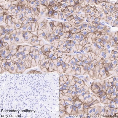
Immunohistochemical analysis of paraffin-embedded human thyroid cancer tissue labeling EpCAM with ab223582 at 1/500 dilution, followed by Goat Anti-Rabbit IgG H&L (HRP) Ready to use. Membranous staining on human thyroid cancer is observed (PMID: 15637741). Counter stained with Hematoxylin.
Secondary antibody only control: Used PBS instead of primary antibody, secondary antibody is Goat Anti-Rabbit IgG H&L (HRP) Ready to use.
Perform heat mediated antigen retrieval with Tris/EDTA buffer pH 9.0 before commencing with IHC staining protocol.
-
 Immunohistochemistry (Formalin/PFA-fixed paraffin-embedded sections) - EpCAM (EPR20532-225, AUA1, EPR20532-222, EPR677(2)) Antibody Sampler Panel - Human, IHC (ab269810)
IHC image of ab20160 staining in human breast carcinoma formalin fixed paraffin embedded tissue section, performed on a Leica BondTM system using the standard protocol F.
The section was pre-treated using heat mediated antigen retrieval with sodium citrate buffer (pH 6, epitope retrieval solution 1) for 20 mins. The section was then incubated with ab20160, 10µg/ml, for 15 mins at room temperature and detected using an HRP conjugated compact polymer system. DAB was used as the chromogen. The section was then counterstained with hematoxylin and mounted with DPX.
For other IHC staining systems (automated and non-automated) customers should optimize variable parameters such as antigen retrieval conditions, primary antibody concentration and antibody incubation times. -
 Immunohistochemistry (Formalin/PFA-fixed paraffin-embedded sections) - EpCAM (EPR20532-225, AUA1, EPR20532-222, EPR677(2)) Antibody Sampler Panel - Human, IHC (ab269810)
Immunohistochemical analysis of paraffin-embedded human colon tissue labeling EpCAM with ab213500 at 1/16000 dilution, followed by Goat Anti-Rabbit IgG H&L (HRP) (ab97051) at 1/500 dilution.
Membranous staining on human colon is observed [PMID: 15637741].
Counter stained with Hematoxylin.
Secondary antibody only control: Used PBS instead of primary antibody, secondary antibody is Goat Anti-Rabbit IgG H&L (HRP) (ab97051) at 1/500 dilution.
Perform heat mediated antigen retrieval with Tris/EDTA buffer pH 9.0 before commencing with IHC staining protocol.
-
 Immunohistochemistry (Formalin/PFA-fixed paraffin-embedded sections) - EpCAM (EPR20532-225, AUA1, EPR20532-222, EPR677(2)) Antibody Sampler Panel - Human, IHC (ab269810)
Immunohistochemical analysis of paraffin-embedded human endometrium cancer tissue labeling EpCAM with ab213500 at 1/16000 dilution, followed by Goat Anti-Rabbit IgG H&L (HRP) (ab97051) at 1/500 dilution.
Membranous staining on tumor cells of human endometrium cancer is observed [PMID: 15637741].
Counter stained with Hematoxylin.
Secondary antibody only control: Used PBS instead of primary antibody, secondary antibody is Goat Anti-Rabbit IgG H&L (HRP) (ab97051) at 1/500 dilution.
Perform heat mediated antigen retrieval with Tris/EDTA buffer pH 9.0 before commencing with IHC staining protocol.
-
 Immunohistochemistry (Formalin/PFA-fixed paraffin-embedded sections) - EpCAM (EPR20532-225, AUA1, EPR20532-222, EPR677(2)) Antibody Sampler Panel - Human, IHC (ab269810)
ab124825, at 1/100 dilution, staining EpCAM in paraffin-embedded Human colon adenocarcinoma tissue by Immunohistochemistry.
Perform heat mediated antigen retrieval before commencing with IHC staining protocol.
-
 Immunohistochemistry (Formalin/PFA-fixed paraffin-embedded sections) - EpCAM (EPR20532-225, AUA1, EPR20532-222, EPR677(2)) Antibody Sampler Panel - Human, IHC (ab269810)
ab124825, at 1/100 dilution, staining EpCAM in paraffin-embedded Human colon tissue by Immunohistochemistry.
Perform heat mediated antigen retrieval before commencing with IHC staining protocol.
-
 Immunohistochemistry (Formalin/PFA-fixed paraffin-embedded sections) - EpCAM (EPR20532-225, AUA1, EPR20532-222, EPR677(2)) Antibody Sampler Panel - Human, IHC (ab269810)
ab124825, at 1/100 dilution, staining EpCAM in paraffin-embedded Human endometrial adenocarcinoma tissue by Immunohistochemistry.
Perform heat mediated antigen retrieval before commencing with IHC staining protocol.
-


