Anti-Wnt6 antibody (ab50030)
Key features and details
- Rabbit polyclonal to Wnt6
- Suitable for: WB, ICC/IF
- Reacts with: Human
- Isotype: IgG
Overview
-
Product name
Anti-Wnt6 antibody
See all Wnt6 primary antibodies -
Description
Rabbit polyclonal to Wnt6 -
Host species
Rabbit -
Tested applications
Suitable for: WB, ICC/IFmore details -
Species reactivity
Reacts with: Human
Predicted to work with: Mouse
-
Immunogen
Synthetic peptide corresponding to Human Wnt6 aa 250-350 conjugated to keyhole limpet haemocyanin.
(Peptide available asab50029) -
Positive control
- This antibody gave a positive signal in the following whole cell lysates: HeLa (Human epithelial carcinoma cell line); Jurkat (Human T cell lymphoblast-like cell line); A431 (Human epithelial carcinoma cell line); HEK293 (Human embryonic kidney cell line); HepG2 (Human hepatocellular liver carcinoma cell line); MCF7 (Human breast adenocarcinoma cell line); SHSY-5Y (Human neuroblastoma cell line); U2OS (Human osteosarcoma cell line)
Properties
-
Form
Liquid -
Storage instructions
Shipped at 4°C. Store at +4°C short term (1-2 weeks). Upon delivery aliquot. Store at -20°C or -80°C. Avoid freeze / thaw cycle. -
Storage buffer
pH: 7.40
Preservative: 0.02% Sodium azide
Constituent: PBS
Batches of this product that have a concentration Concentration information loading...
Purity
Immunogen affinity purifiedClonality
PolyclonalIsotype
IgGResearch areas
Associated products
-
Compatible Secondaries
-
Isotype control
-
Recombinant Protein
Applications
Our Abpromise guarantee covers the use of ab50030 in the following tested applications.
The application notes include recommended starting dilutions; optimal dilutions/concentrations should be determined by the end user.
Application Abreviews Notes WB Use a concentration of 1 µg/ml. Predicted molecular weight: 40 kDa. ICC/IF Use at an assay dependent concentration. Target
-
Function
Ligand for members of the frizzled family of seven transmembrane receptors. Probable developmental protein. May be a signaling molecule which affects the development of discrete regions of tissues. Is likely to signal over only few cell diameters. -
Sequence similarities
Belongs to the Wnt family. -
Post-translational
modificationsPalmitoylation at Ser-228 is required for efficient binding to frizzled receptors. It is also required for subsequent palmitoylation at Cys-76. Palmitoylation is necessary for proper trafficking to cell surface. -
Cellular localization
Secreted > extracellular space > extracellular matrix. - Information by UniProt
-
Database links
- Entrez Gene: 7475 Human
- Entrez Gene: 22420 Mouse
- Omim: 604663 Human
- SwissProt: Q9Y6F9 Human
- SwissProt: P22727 Mouse
- Unigene: 29764 Human
- Unigene: 268282 Mouse
-
Alternative names
- Protein Wnt 6 antibody
- Protein Wnt-6 antibody
- Protein Wnt-6 precursor antibody
see all
Images
-
All lanes : Anti-Wnt6 antibody (ab50030) at 1 µg/ml
Lane 1 : HeLa (Human epithelial carcinoma cell line) Whole Cell Lysate
Lane 2 : Jurkat (Human T cell lymphoblast-like cell line) Whole Cell Lysate
Lane 3 : A431 (Human epithelial carcinoma cell line) Whole Cell Lysate
Lane 4 : HEK293 Human embryonic kidney cell line Whole Cell Lysate
Lane 5 : HepG2 (Human hepatocellular liver carcinoma cell line) Whole Cell Lysate
Lane 6 : MCF7 (Human breast adenocarcinoma cell line) Whole Cell Lysate
Lane 7 : SHSY-5Y (Human neuroblastoma cell line) Whole Cell Lysate
Lane 8 : U2OS (Human osteosarcoma cell line) Whole Cell Lysate
Lysates/proteins at 10 µg per lane.
Secondary
All lanes : IRDye 680 Conjugated Goat Anti-Rabbit IgG (H+L) at 1/10000 dilution
Performed under reducing conditions.
Predicted band size: 40 kDa
Observed band size: 55 kDa why is the actual band size different from the predicted?
Wnt6 has a predicted molecular weight of 40 kDa; however it has a number of potential glycosylation sites which may affect the migration of the protein (SwissProt data). Abcam currently sell two antibodies to this target, ab49946 which detects a band at 78 kDa and ab50030 which detects a band at 55 kDa. Abcam welcomes customer feedback and would appreciate any comments regarding this product and the data presented above. -
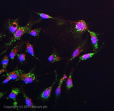
ICC/IF image of ab50030 stained HepG2 cells. The cells were 4% PFA fixed (10 min) and then incubated in 1%BSA / 10% normal goat serum / 0.3M glycine in 0.1% PBS-Tween for 1h to permeabilise the cells and block non-specific protein-protein interactions. The cells were then incubated with the antibody (ab50030, 1µg/ml) overnight at +4°C. The secondary antibody (green) was ab96899 Dylight 488 goat anti-rabbit IgG (H+L) used at a 1/250 dilution for 1h. Alexa Fluor® 594 WGA was used to label plasma membranes (red) at a 1/200 dilution for 1h. DAPI was used to stain the cell nuclei (blue) at a concentration of 1.43µM.
Protocols
Datasheets and documents
References (9)
ab50030 has been referenced in 9 publications.
- Gonçalves CS et al. A novel molecular link between HOXA9 and WNT6 in glioblastoma identifies a subgroup of patients with particular poor prognosis. Mol Oncol 14:1224-1241 (2020). PubMed: 31923345
- Peng J et al. High WNT6 expression indicates unfavorable survival outcome for patients with colorectal liver metastasis after liver resection. J Cancer 10:2619-2627 (2019). PubMed: 31258769
- Zheng XL & Yu HG Wnt6 contributes tumorigenesis and development of colon cancer via its effects on cell proliferation, apoptosis, cell-cycle and migration. Oncol Lett 16:1163-1172 (2018). PubMed: 29963191
- Gonçalves CS et al. WNT6 is a novel oncogenic prognostic biomarker in human glioblastoma. Theranostics 8:4805-4823 (2018). PubMed: 30279739
- Pope C et al. The role of H19, a long non-coding RNA, in mouse liver postnatal maturation. PLoS One 12:e0187557 (2017). PubMed: 29099871
- Zhang L et al. Increased WNT6 expression in tumor cells predicts unfavorable survival in esophageal squamous cell carcinoma patients. Int J Clin Exp Pathol 8:11421-7 (2015). PubMed: 26617869
- Richards MH et al. Porcupine is not required for the production of the majority of Wnts from primary human astrocytes and CD8+ T cells. PLoS One 9:e92159 (2014). WB . PubMed: 24647048
- Yuan G et al. WNT6 is a novel target gene of caveolin-1 promoting chemoresistance to epirubicin in human gastric cancer cells. Oncogene : (2012). WB, IHC . PubMed: 22370641
- Wang C et al. Effect of Wnt6 on human dental papilla cells in vitro. J Endod 36:238-43 (2010). WB ; Human . PubMed: 20113781
Images
-
All lanes : Anti-Wnt6 antibody (ab50030) at 1 µg/ml
Lane 1 : HeLa (Human epithelial carcinoma cell line) Whole Cell Lysate
Lane 2 : Jurkat (Human T cell lymphoblast-like cell line) Whole Cell Lysate
Lane 3 : A431 (Human epithelial carcinoma cell line) Whole Cell Lysate
Lane 4 : HEK293 Human embryonic kidney cell line Whole Cell Lysate
Lane 5 : HepG2 (Human hepatocellular liver carcinoma cell line) Whole Cell Lysate
Lane 6 : MCF7 (Human breast adenocarcinoma cell line) Whole Cell Lysate
Lane 7 : SHSY-5Y (Human neuroblastoma cell line) Whole Cell Lysate
Lane 8 : U2OS (Human osteosarcoma cell line) Whole Cell Lysate
Lysates/proteins at 10 µg per lane.
Secondary
All lanes : IRDye 680 Conjugated Goat Anti-Rabbit IgG (H+L) at 1/10000 dilution
Performed under reducing conditions.
Predicted band size: 40 kDa
Observed band size: 55 kDa why is the actual band size different from the predicted?
Wnt6 has a predicted molecular weight of 40 kDa; however it has a number of potential glycosylation sites which may affect the migration of the protein (SwissProt data). Abcam currently sell two antibodies to this target, ab49946 which detects a band at 78 kDa and ab50030 which detects a band at 55 kDa. Abcam welcomes customer feedback and would appreciate any comments regarding this product and the data presented above. -
ICC/IF image of ab50030 stained HepG2 cells. The cells were 4% PFA fixed (10 min) and then incubated in 1%BSA / 10% normal goat serum / 0.3M glycine in 0.1% PBS-Tween for 1h to permeabilise the cells and block non-specific protein-protein interactions. The cells were then incubated with the antibody (ab50030, 1µg/ml) overnight at +4°C. The secondary antibody (green) was ab96899 Dylight 488 goat anti-rabbit IgG (H+L) used at a 1/250 dilution for 1h. Alexa Fluor® 594 WGA was used to label plasma membranes (red) at a 1/200 dilution for 1h. DAPI was used to stain the cell nuclei (blue) at a concentration of 1.43µM.


