Anti-UCP1 antibody (ab23841)
Key features and details
- Rabbit polyclonal to UCP1
- Suitable for: WB
- Reacts with: Mouse, Rat
- Isotype: IgG
Overview
-
Product name
Anti-UCP1 antibody
See all UCP1 primary antibodies -
Description
Rabbit polyclonal to UCP1 -
Host species
Rabbit -
Tested applications
Suitable for: WBmore details -
Species reactivity
Reacts with: Mouse, Rat
Predicted to work with: Hamster, Dog, Human, Chimpanzee
-
Immunogen
Synthetic peptide corresponding to Human UCP1 aa 100-200 conjugated to keyhole limpet haemocyanin.
(Peptide available asab24282) -
General notes
Reproducibility is key to advancing scientific discovery and accelerating scientists’ next breakthrough.
Abcam is leading the way with our range of recombinant antibodies, knockout-validated antibodies and knockout cell lines, all of which support improved reproducibility.
We are also planning to innovate the way in which we present recommended applications and species on our product datasheets, so that only applications & species that have been tested in our own labs, our suppliers or by selected trusted collaborators are covered by our Abpromise™ guarantee.
In preparation for this, we have started to update the applications & species that this product is Abpromise guaranteed for.
We are also updating the applications & species that this product has been “predicted to work with,” however this information is not covered by our Abpromise guarantee.
Applications & species from publications and Abreviews that have not been tested in our own labs or in those of our suppliers are not covered by the Abpromise guarantee.
Please check that this product meets your needs before purchasing. If you have any questions, special requirements or concerns, please send us an inquiry and/or contact our Support team ahead of purchase. Recommended alternatives for this product can be found below, as well as customer reviews and Q&As.
Properties
-
Form
Liquid -
Storage instructions
Shipped at 4°C. Store at +4°C short term (1-2 weeks). Upon delivery aliquot. Store at -20°C or -80°C. Avoid freeze / thaw cycle. -
Storage buffer
pH: 7.40
Preservative: 0.02% Sodium azide
Constituent: PBS
Batches of this product that have a concentration Concentration information loading...
Purity
Immunogen affinity purifiedClonality
PolyclonalIsotype
IgGResearch areas
Associated products
-
Compatible Secondaries
-
Immunizing Peptide (Blocking)
-
Isotype control
-
Recombinant Protein
Applications
Our Abpromise guarantee covers the use of ab23841 in the following tested applications.
The application notes include recommended starting dilutions; optimal dilutions/concentrations should be determined by the end user.
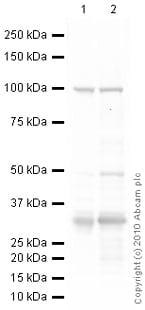
Application Abreviews Notes WB Use a concentration of 1 µg/ml. Detects a band of approximately 33 kDa (predicted molecular weight: 33 kDa). Target
-
Function
UCP are mitochondrial transporter proteins that create proton leaks across the inner mitochondrial membrane, thus uncoupling oxidative phosphorylation from ATP synthesis. As a result, energy is dissipated in the form of heat. -
Tissue specificity
Brown adipose tissue. -
Sequence similarities
Belongs to the mitochondrial carrier family.
Contains 3 Solcar repeats. -
Cellular localization
Mitochondrion inner membrane. - Information by UniProt
-
Database links
- Entrez Gene: 7350 Human
- Entrez Gene: 22227 Mouse
- Entrez Gene: 24860 Rat
- Omim: 113730 Human
- SwissProt: P25874 Human
- SwissProt: P12242 Mouse
- SwissProt: P04633 Rat
- Unigene: 249211 Human
see all -
Form
UCP1 is preferentially expressed in brown adipose tissue -
Alternative names
- mitochondrial brown fat uncoupling protein antibody
- Mitochondrial brown fat uncoupling protein 1 antibody
- SLC25A7 antibody
see all
Images
-
All lanes : Anti-UCP1 antibody (ab23841) at 1 µg/ml
Lane 1 : Adult Mouse Brown Adipose Tissue Lysate
Lane 2 : Adult Rat Brown Adipose Tissue Lysate
Lysates/proteins at 20 µg per lane.
Secondary
All lanes : Goat polyclonal to Rabbit IgG - H&L - Pre-Adsorbed (HRP) at 1/3000 dilution
Performed under reducing conditions.
Predicted band size: 33 kDa
Observed band size: 33 kDa
Additional bands at: 100 kDa, 50 kDa. We are unsure as to the identity of these extra bands.
Exposure time: 30 seconds -
 Immunohistochemistry - Anti-UCP1 antibody (ab23841)Image from Liu W et al., PLoS Genet. 2006;8:11958-63. Fig 5.; doi: 10.1371/journal.pgen.1003626 Reproduced under the Creative Commons license http://creativecommons.org/licenses/by/4.0/
ab23841 staining UCP1 in serial sections of white fat from mice by immunohistochemistry. Serial sections were de-paraffinized, rehydrated through xylene, ethanol and water; antigen retrieval was by heat mediation in a citrate buffer. Samples were incubated with primary antibody (1/200 in blocking buffer) for 60 minutes. A HRP-conjugated anti-rabbit IgG was used as the secondary antibody.
References (93)
ab23841 has been referenced in 93 publications.
- Johnson JM et al. Alternative splicing of UCP1 by non-cell-autonomous action of PEMT. Mol Metab 31:55-66 (2020). PubMed: 31918922
- Shamsi F et al. FGF6 and FGF9 regulate UCP1 expression independent of brown adipogenesis. Nat Commun 11:1421 (2020). PubMed: 32184391
- Wei G et al. Indirubin, a small molecular deriving from connectivity map (CMAP) screening, ameliorates obesity-induced metabolic dysfunction by enhancing brown adipose thermogenesis and white adipose browning. Nutr Metab (Lond) 17:21 (2020). PubMed: 32190098
- Lee K et al. Gomisin N from Schisandra chinensis Ameliorates Lipid Accumulation and Induces a Brown Fat-Like Phenotype through AMP-Activated Protein Kinase in 3T3-L1 Adipocytes. Int J Mol Sci 21:N/A (2020). PubMed: 32245100
- Tajima K et al. Mitochondrial lipoylation integrates age-associated decline in brown fat thermogenesis. Nat Metab 1:886-898 (2019). PubMed: 32313871
Images
-
All lanes : Anti-UCP1 antibody (ab23841) at 1 µg/ml
Lane 1 : Adult Mouse Brown Adipose Tissue Lysate
Lane 2 : Adult Rat Brown Adipose Tissue Lysate
Lysates/proteins at 20 µg per lane.
Secondary
All lanes : Goat polyclonal to Rabbit IgG - H&L - Pre-Adsorbed (HRP) at 1/3000 dilution
Performed under reducing conditions.
Predicted band size: 33 kDa
Observed band size: 33 kDa
Additional bands at: 100 kDa, 50 kDa. We are unsure as to the identity of these extra bands.
Exposure time: 30 seconds
-
 Immunohistochemistry - Anti-UCP1 antibody (ab23841) Image from Liu W et al., PLoS Genet. 2006;8:11958-63. Fig 5.; doi: 10.1371/journal.pgen.1003626 Reproduced under the Creative Commons license http://creativecommons.org/licenses/by/4.0/
ab23841 staining UCP1 in serial sections of white fat from mice by immunohistochemistry. Serial sections were de-paraffinized, rehydrated through xylene, ethanol and water; antigen retrieval was by heat mediation in a citrate buffer. Samples were incubated with primary antibody (1/200 in blocking buffer) for 60 minutes. A HRP-conjugated anti-rabbit IgG was used as the secondary antibody.


