Anti-TLR4 antibody (ab13556)
Key features and details
- Rabbit polyclonal to TLR4
- Suitable for: WB, IHC-P, IHC-Fr, Flow Cyt
- Reacts with: Mouse, Human, Recombinant fragment
- Isotype: IgG
Overview
-
Product name
Anti-TLR4 antibody
See all TLR4 primary antibodies -
Description
Rabbit polyclonal to TLR4 -
Host species
Rabbit -
Specificity
TLR4 expression levels and cleavage or degradation products can vary between different cell and tissue samples. Customers have observed this variability in WB band size and our laboratory has confirmed this variability as well observing lower molecular weight cleavage and degradation products and in some samples a lack of the full length TLR4 band. The TLR4 cleavage and degradation products and potential lack of full length TLR4 are well documented in the literature, including PMID 16885150 and 22927440. We recommend running a positive control human intestine tissue lysate. We have obtained both positive and negative feedback from researchers using this antibody with rat samples (see Abreviews). Due to the inconsistency, we have removed rat as a guaranteed species and welcome any further feedback from researchers using this antibody. -
Tested Applications & Species
See all applications and species dataApplication Species Flow Cyt HumanIHC-P MouseWB MouseRecombinant fragment -
Immunogen
Synthetic peptide corresponding to Human TLR4 aa 420-435.
Sequence:GLEQLEHLDFQ HSNLK
Database link: O00206
Properties
-
Form
Liquid -
Storage instructions
Shipped at 4°C. Store at +4°C short term (1-2 weeks). Upon delivery aliquot. Store at -20°C or -80°C. Avoid freeze / thaw cycle. -
Storage buffer
Preservative: 0.05% Sodium azide
Constituents: 99% PBS, 0.05% BSA -
 Concentration information loading...
 -
Purity
Protein G purified -
Clonality
Polyclonal -
Isotype
IgG -
Research areas
Images
-
Anti-TLR4 antibody (ab13556) at 1/1000 dilution + partial recombinant mouse TLR4 protein, 100 ng
-
Anti-TLR4 antibody (ab13556) at 1/500 dilution + TLR4 transfected Baculovirus-Insect whole cell lysate at 10 µg
Secondary
Anti-Rabbit HRP conjugate at 1/2000 dilution
Exposure time: 6 minutes
-
ab13556 at a 1/100 dilution staining TLR4 in mouse spleen tissue section by Immunohistochemistry (Formalin/PFA-fixed paraffin-embedded sections).
-
ab13556 at a 1/100 dilution staining TLR4 in mouse spleen tissue section by Immunohistochemistry (Formalin/PFA-fixed paraffin-embedded sections).
-
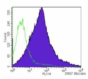
 Flow Cytometry - Anti-TLR4 antibody (ab13556) This image is courtesy of an Abreview submitted by Dr alexandre garin
ab13556 diluted 1/100 detecting TLR4 transfected mouse CHO cells by flow cytometry. The cells were prepared by treatment with collagenase and incubated with the primary antibody for 1 hour at 22°C. An Alexa Fluor® 488 goat anti-rabbit was used as the secondary antibody. Cells gated on live.
-
ab13556 at 1/100000 dilution (12 hrs at 4degC) staining TLR4 in mouse colitis colon tissue section by Immunohistochemistry (Formalin-fixed tissue sections). A biotin Goat Anti-Rabbit secondary was used at 1/2000 for 1 hour at RT. Counterstain: Methyl Green at 200µL for 2 mins at RT. Localization: Inflammatory cells.
-
Flow cytometry analysis of THP-1 (Human monocytic leukemia cell line). Cells were fixed with 2% formaldehyde for 10 minutes at room temperature. ab13556 was used at 2 µg/106 cells for 60 minutes at 37°C (green). Goat Anti- Rabbit Dylight 488 was used as a secondary antibody at 1/200 dilution for 40 minutes at 37°C. Isotype control was Rabbit IgG under the same conditions (blue).


